Log in    RU  


Main page :: Forum :: Ultrasound cases - Sonograms, cine-loops etc. ::

Area with hyperechoic dot inclusions


Tags: Thyroid gland sonography, GE Logiq 400 MD, Clinical report, Pediatric

   Posts
 
  Area with hyperechoic dot inclusion...
#1

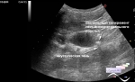
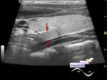
    A 13-year-old teenager was sent to the public clinic for preventive ultrasound of the thyroid gland.

    On ultrasound in the right lobe, hyperechoic dot inclusions are visualized (differential diagnosis: microcalcifications, thymus ectopia, etc.), an isoechoic node without a clear contour cannot be excluded.


 Random 
Admin room - Who are Sonologists?

Ultrasound cases - Discharge from the navel
    

Ultrasound cases - Earrings and consequences
    

Ultrasound cases - Suspicion of gastritis or renal colic
    

Ultrasound cases - Mickey Mouse sign
    

 Ad 


 Dictionary 
ESD

MPAP

HR

VTI

LVPWd

Ved

GS

Doppler angle

PSAX

LVLs


 Calculators 
Game: Sonographer certification

Gestational age by Ultrasound

Myocardial mass

Body surface area

Estimated fetal weight by Ultrasound


 Similar topics 
Ultrasound cases - Tirads 5 multinodular
    

Ultrasound cases - Pediatric Tirads 5
    

Ultrasound cases - Tirads 5
    

Ultrasound cases - Thyroiditis 2
    

Ultrasound cases - Diffuse-focal thyroid changes, parathyroid glands adenomas
    

Ultrasound cases - Accessory Thyroid Lobe
    

Ultrasound cases - Thyroiditis
    

Ultrasound cases - Hypoplasia of the left thyroid lobe
    

Ultrasound cases - Cystic-solid volume of the thyroid gland
    

Ultrasound cases - Hyperparathyroidism?