Log in    RU  


Main page :: Forum :: Ultrasound cases - Sonograms, cine-loops etc. ::

Renal pelvis valve-stone


Tags: Urinary tract sonography, Esaote MyLab 70, Clinical report, Pediatric

   Posts
 
  Renal pelvis valve-stone
#1

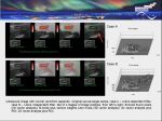
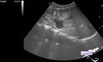
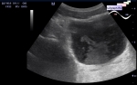
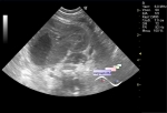
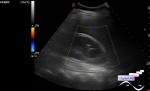
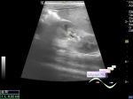
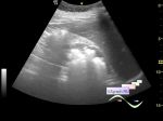
    A 10-year-old child with hematuria and suspicion of acute cystitis or trauma was admitted via the ambulance to the Children's City Clinical Hospital and was sent urgently for an urinary tract ultrasound scan from the ED.

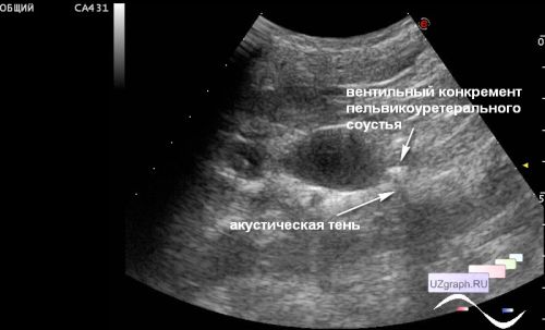
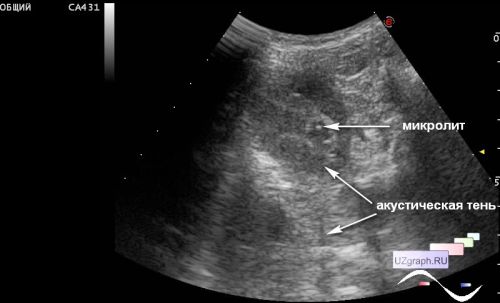
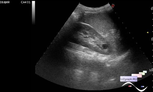
    On ultrasound, the right kidney with diffuse dilated collecting system, pelvis up to 1.5 cm, calixes up to 7 mm. In the area of ​​the pyeloureteral junction, a stone up to 7 mm with an acoustic shadow is visualized, parapelvic ureter up to 5 μμ - a renal pelvis valve-stone. The calixes contain multiple echo-positive inclusions up to 2 mm (kidney microlithiasis). In the upper third of the left kidney, a cyst-like structure up to 9 mm, presumably a single dilated calix (Fraley syndrome).


 Random 
Ultrasound cases - Beta-thalassemia and vermiform appendix
    

Ultrasound cases - After anus atresia
    

Ultrasound cases - Pneumatosis intestinalis, intussusception etc.
    

Ultrasound cases - Linea alba hernia plus
    

Ultrasound cases - Gallbladder, saw sign
    

 Dictionary 
VTI

SF

RAP

TV

Ved

CRL

Aliasing doppler artifact

HC

Doppler angle

LVESV


 Calculators 
TI-RADS

Myocardial mass

Volume (ellipse formula)

Teicholz systolic cardiac function

Qp:Qs


 Similar topics 
Ultrasound cases - Autosomal recessive polycystic kidney disease (ARPKD)
    

Ultrasound cases - Bladder sludge
    

Ultrasound cases - Bright kidneys
    

Ultrasound cases - Ileostoma stenosis and nephropathy
    

Ultrasound cases - Renal pelvis clot
    

Ultrasound cases - Condition after treatment of a kidney cyst
    

Ultrasound cases - Hydronephrotic transformation & polypous bladder wall
    

Ultrasound cases - Pyelitis, cystitis
    

Ultrasound cases - Lesion in the projection of the adrenal gland 2
    

Ultrasound cases - Hyperechoic renal sinus with acoustic shadow