Log in    RU  


Main page :: Forum :: Ultrasound cases - Sonograms, cine-loops etc. ::

Air bronchogram


Tags: Chest sonography, GE Logiq Book XP, Clinical report, Pediatric

   Posts
 
  Air bronchogram
#1

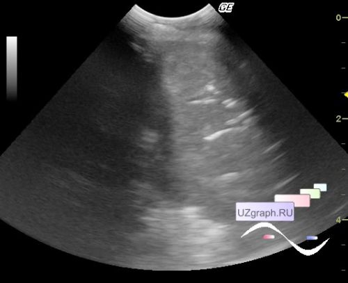
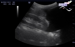
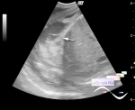
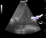
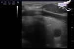
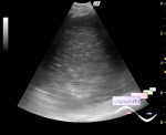
    A newborn in the ICU of the Children's City Clinical Hospital after surgery on the esophagus (atresia), an ultrasound of the pleural cavities was prescribed for fluid.

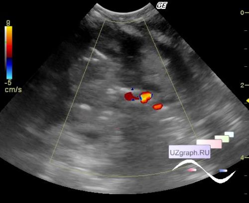
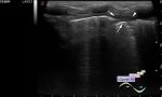
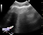
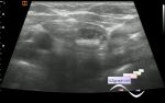
    At Ultrasound in the left half of the chest visualizes a parenchymal structure with blood flow at CFM and tree-like hyperechoic structures (bronchial tree with air - air bronchogram) along which the movement of hyperechoic microinclusions (air bubbles) is periodically observed.

    Dif. diagnosis: postoperative lung atelectasis, etc.


 Random 
Ultrasound cases - Hydrocele and rete
    

Ultrasound cases - Hirschsprung's
disease after surgery


Ultrasound cases - Duodenal atresia
    

Ultrasound cases - Scrotum hematocele
    

Ultrasound cases - Good vs Evil - thyroid 3
    

 Dictionary 
PSV

LVEDV

EDD

Ved

LVESD

BSA

DPD

CFM

GA

B-mode


 Calculators 
Thyroid volume

Volume (ellipse formula)

Type of an infant hip joint

Teicholz systolic cardiac function

Pulmonary artery pressure


 Similar topics 
Ultrasound cases - Chylopericardium, lung atelectasis, cardiomegaly, ascites
    

Ultrasound cases - Diaphragmatic hernia, condition after surgery
    

Ultrasound cases - Hydrothorax
    

Ultrasound cases - One and a half kilograms of happiness
    

Ultrasound cases - Mirror artifact vs collapsed lung
    

Ultrasound cases - Persistent thymus or not
    

Ultrasound cases - Postoperative hydrothorax with sludge
    

Ultrasound cases - Pneumonia and hydrothorax on the left
    

Ultrasound cases - Diaphragmatic hernia, ectopic spleen & air...
    

Ultrasound cases - Diffusive-focal changes of the thymus