Diaphragmatic hernia, ectopic spleen & air...
Tags: Chest sonography, Abdomen sonography, Cardiac sonography(Echocardiography), GE Logiq Book XP, Images, Video, Clinical report, Pediatric
| Posts | |||
| Diaphragmatic hernia, ectopic spleen ... | #1 |
| |||||
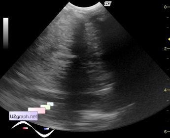
:: file 1 ::
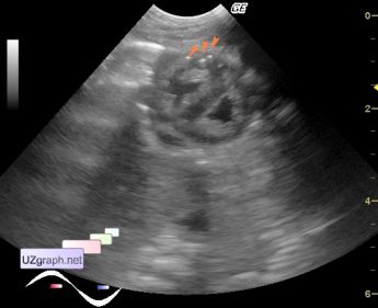
 :: file 2 ::
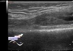
 :: file 3 ::
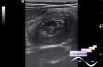
 :: file 4 ::
 :: file 5 ::
 :: file 6 ::
 :: file 7 :: | |||||