Log in    RU  


Main page :: Forum :: Ultrasound cases - Sonograms, cine-loops etc. ::

Hemangioma of the chin area


Tags: Soft tissues sonography, Images, Video, Clinical report, Esaote MyLab 70, Pediatric

   Posts
 
  Hemangioma of the chin area
#1

    Child 2 years with hemangioma of chin area, previously has puncture in order to verify the mass, at this time came to doctor with complains on an increasing mass in a few times over the last few days, sent for an soft tissue ultrasound.

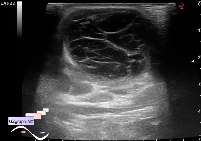
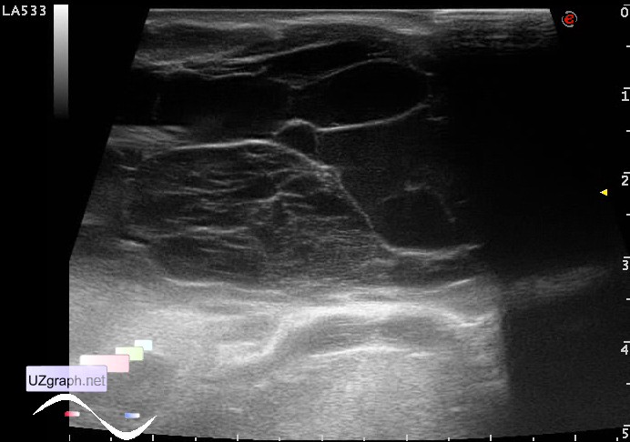
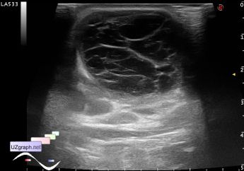
    Visually mass occupies the entire chin, size of mass about 10 x 5 cm or more.

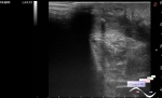
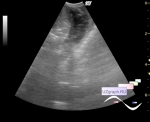
    On ultrasound the mass has the structure as encysted fluid type - an / hypoechoic with hyperechoic septums.

    CFM failed to appreciate - the child was constantly crying.

    external link

:: attachments(3) ::

:: file 1 ::

:: file 2 ::

:: file 3 ::


 Random 
Ultrasound cases - Thyroid lesion
    

Ultrasound cases - Recurrent shin trauma
    

Ultrasound cases - 1 kilogram of happiness
    

Scientific - Lecture about LUS at COVID-19
    

Ultrasound cases - Abdominal cryptorchidism
    

 Dictionary 
EFW

IVC

MC

FNA

VTI

CFM

PW

Anatomical M-mode

PD

EF


 Calculators 
Volume (ellipse formula)

Pulmonary artery pressure

Type of an infant hip joint

Pressure gradient

Gestational age by Ultrasound


 Similar topics 
Ultrasound cases - Penetrating injury of the femur
    

Ultrasound cases - Cephalohematoma
    

Ultrasound cases - Urachus fistula
    

Ultrasound cases - Cranial mass
    

Ultrasound cases - Pilonidal coccyx cyst
    

Ultrasound cases - Palm fistula, in search of pencil lead
    

Ultrasound cases - Postoperative abdominal wall fistula
    

Ultrasound cases - Nucks cyst
    

Ultrasound cases - Paraproctitis
    

Ultrasound cases - Submandibular area lesion