Log in    RU  


Main page :: Forum :: Ultrasound cases - Sonograms, cine-loops etc. ::

Lesion of the distal third of the right femur


Tags: Musculoskeletal sonography, Soft tissues sonography, Video, Clinical report, Esaote MyLab 70, Pediatric

   Posts
 
  Lesion of the distal third of the r...
#1

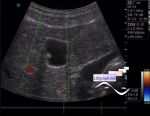
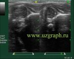
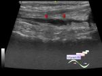
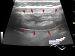
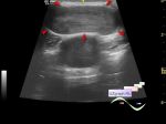
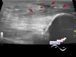
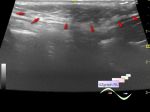
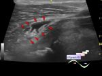
    Child 5 years old with suspected arthritis of the knee.

    Previously, by the patient' s words, was sharply swollen knee, injury denies, previous ultrasound found fluid.

    At US in the antero-medial distal third of the right femur visualized heteroechoic lesion type of complex cyst with thickened wall, uneven content, with peripheral blood flow at CFM.

    The narrow isthmus of lesion is lost in the projection of the medial meniscus, the approximate size of the lesion is 4x2,5x6 cm (cyst of the medial meniscus with rupture? else?)

    In the projection of the same side popliteal fossa visualized enlarged lymph node, with a blood flow at CFM, size of up to 8mm.

    external link

:: attachments(1) ::

:: file 1 ::

  08:31 18-11-2019
 
#2

    Possible hemarthrosis


 Random 
Ultrasound cases - Ovarian apoplexy
    

Ultrasound cases - Non visualization of testicles
    

Ultrasound cases - Another polyp of the gallbladder
    

Ultrasound cases - Subluxation of the hip joint (type III Graf)
    

Ultrasound cases - Accessory thymic lobe
    

 Dictionary 
EFW

B-mode

EDD

Anatomical M-mode

VTI

RVOT

PASP

BPD

LVPWd

BSA


 Calculators 
Gestational age by Ultrasound

Pressure gradient

TI-RADS

Thyroid volume

Teicholz systolic cardiac function


 Similar topics 
Ultrasound cases - Penetrating injury of the femur
    

Ultrasound cases - Cephalohematoma
    

Ultrasound cases - Cranial mass
    

Ultrasound cases - Recurrent shin trauma
    

Ultrasound cases - Knee joint synovitis
    

Ultrasound cases - Thickening and heterogeneity of the intercostal space soft tissues
    

Ultrasound cases - Chest wall lesion
    

Ultrasound cases - Hockey Elbow
    

Ultrasound cases - Combination of left shoulder lesions
    

Ultrasound cases - Baker's cyst and intramuscular hematoma