Log in    RU  


Main page :: Forum :: Ultrasound cases - Sonograms, cine-loops etc. ::

Chest wall lesion


Tags: Soft tissues sonography, Musculoskeletal sonography, GE Logiq E, Clinical report, Pediatric

   Posts
 
  Chest wall lesion
#1

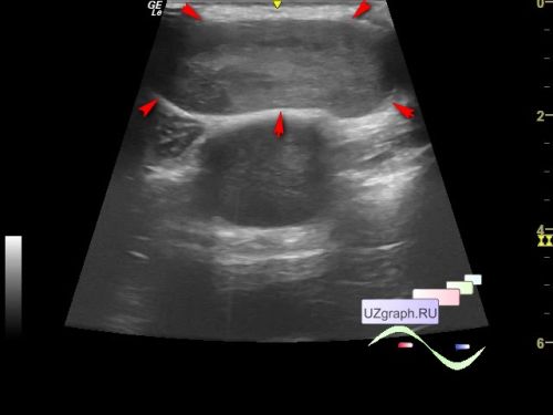
    A 16-year-old teenager was referred for a chest soft tissues ultrasound due to the lesion on the lateral surface of the chest, with which he has been observed, if I understood everything correctly, for several years, according to his words, ultrasound and X-rays had already been performed earlier (MRI was not performed), and doctors called this lesion a cyst.

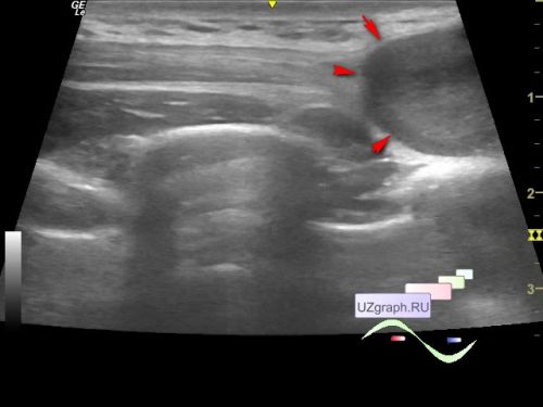
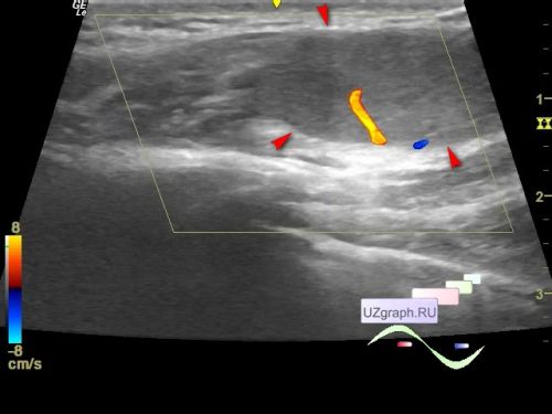
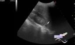
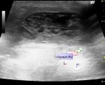
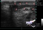
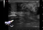
    In the projection of a lesion visible to the naked eye on the right lateral surface of the chest, an intramuscular, heterogeneous, hypoechoic, oval-shaped lesion up to 4 x 4 x 2 cm in size is visualized on ultrasound; at the CFM the blood flow is visualized along the posterior surface of the lesion with arterial type in the spectral mode (differential diagnosis: lipoma, schwannoma, etc.).


 Random 
Ultrasound cases - Bruising/ hematoma/ rupture of the spleen
    

Scientific - ESC Webinar: LUS in COVID-19

Ultrasound cases - Pain in the right iliac region
    

Ultrasound cases - Inflamed mammary cyst(mastitis)
    

Ultrasound cases - Lesion in the projection of the pancreas tail plus
    

 Dictionary 
ESV

BPD

Freeze

TVUS

LVESV

IVC

TI-RADS

CRL

MSD

Qp-Qs


 Calculators 
Myocardial mass

Body surface area

Qp:Qs

Resistive index

TI-RADS


 Similar topics 
Ultrasound cases - Penetrating injury of the femur
    

Ultrasound cases - Cephalohematoma
    

Ultrasound cases - Urachus fistula
    

Ultrasound cases - Cranial mass
    

Ultrasound cases - Pilonidal coccyx cyst
    

Ultrasound cases - Palm fistula, in search of pencil lead
    

Ultrasound cases - Postoperative abdominal wall fistula
    

Ultrasound cases - Nucks cyst
    

Ultrasound cases - Paraproctitis
    

Ultrasound cases - Submandibular area lesion