Log in    RU  


Main page :: Forum :: Ultrasound cases - Sonograms, cine-loops etc. ::

Scrotum hematocele


Tags: Scrotum sonography, GE Logiq Book XP, Clinical report, Pediatric

   Posts
 
  Scrotum hematocele
#1

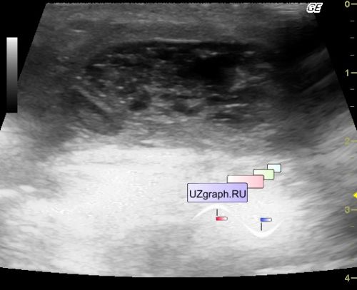
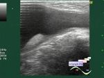
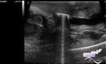
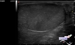
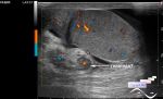
    Infant in the surgical department of the Children's Clinical Hospital after surgery for scrotal cyst removal.

    The scrotum increased again, and therefore an ultrasound scan was assigned.

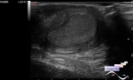
    On ultrasound in the projection of the zone of interest, the formation is of the type of lumped fluid, presumably the scrotum hematoma (hematocele / hemoscrotum).


 Random 
Ultrasound cases - Willis circle at ultrasound
    

Ultrasound cases - Atrioventricular canal
    

Ultrasound cases - Earrings and consequences
    

Ultrasound cases - Thyroid polymorphic lesions
    

On-call room - How to become a sonographer

 Dictionary 
Freeze

CFM

Ved

AC

GA

LVEDV

LVEDD

HC

Gain

TAMEAN


 Calculators 
Type of an infant hip joint

Pressure gradient

Thyroid volume

Estimated fetal weight by Ultrasound

Volume (ellipse formula)


 Similar topics 
Ultrasound cases - Abdominal cryptorchidism
    

Ultrasound cases - Neonatal testicular torsion
    

Ultrasound cases - Fibrinous epididymitis
    

Ultrasound cases - Varicocele in a child
    

Ultrasound cases - Hydrocele (dropsy of the scrotum) in a child
    

Ultrasound cases - Air in the scrotum, post surgery condition
    

Ultrasound cases - Multi-chamber scrotum cyst
    

Ultrasound cases - Diffuse changes of the testicle in a teenager (orchitis)
    

Ultrasound cases - Thickening of the spermatic cord in a teenager
    

Ultrasound cases - Condition after hydatides removal