Log in    RU  


Main page :: Forum :: Ultrasound cases - Sonograms, cine-loops etc. ::

Multi-chamber scrotum cyst


Tags: Scrotum sonography, GE Logiq 400 MD, Clinical report, Pediatric

   Posts
 
  Multi-chamber scrotum cyst
#1

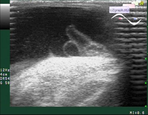
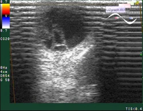
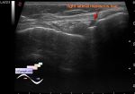
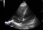
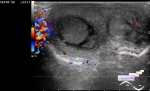
    A child of 3 years old, examined by a surgeon under the screening program in a public clinic, after which he was sent for an urgent ultrasound scan. One hemiscrotum is enlarged on visual inspection. On ultrasound, the testicle from the enlarged side is pressed ro the lower surface of the scrotum by a multi-chamber cyst up to 5 cm in length.


 Random 
Ultrasound cases - Snowball
    

Ultrasound cases - Good vs Evil - thyroid 2
    

Scientific - Playlist Pediatric sonography

Ultrasound cases - Lesion type of torsion of Morgagni's hydatide
    

Ultrasound cases - Only the questions
    

 Dictionary 
LVESV

EDD

Freeze

PG

CRL

VTI

FL

TVUS

LVPWd

IVSd


 Calculators 
Estimated fetal weight by Ultrasound

Body surface area

TI-RADS

Volume (ellipse formula)

Teicholz systolic cardiac function


 Similar topics 
Ultrasound cases - Abdominal cryptorchidism
    

Ultrasound cases - Neonatal testicular torsion
    

Ultrasound cases - Fibrinous epididymitis
    

Ultrasound cases - Scrotum hematocele
    

Ultrasound cases - Varicocele in a child
    

Ultrasound cases - Hydrocele (dropsy of the scrotum) in a child
    

Ultrasound cases - Air in the scrotum, post surgery condition
    

Ultrasound cases - Diffuse changes of the testicle in a teenager (orchitis)
    

Ultrasound cases - Thickening of the spermatic cord in a teenager
    

Ultrasound cases - Condition after hydatides removal