Log in    RU  


Main page :: Forum :: Ultrasound cases - Sonograms, cine-loops etc. ::

Epididymitis


Tags: Scrotum sonography, GE Vivid 3, Images, Video, Clinical report, Adult

   Posts
 
  Epididymitis
#1

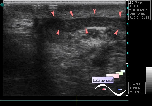
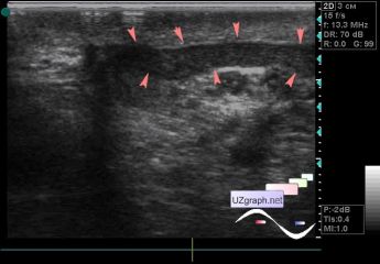
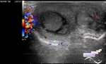
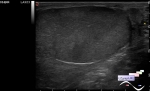
    A 26-year-old patient came to the urologist with complaines about of pain in the scrotum and an increase in testicular density, so directed to an ultrasound.

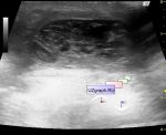
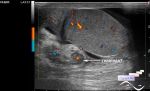
    On ultrasound, the testes are unremarkable, on the side of the complaints, a large hypoechoic region in the epididymis is visualized(epididymitis).

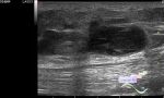
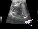
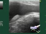
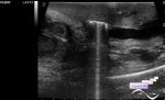
    Ps. Also, you can see in the video the appendages of the epididymis(solid and cystic type).

    external link

:: attachments(3) ::

:: file 1 ::

:: file 2 ::

:: file 3 ::


 Random 
Ultrasound cases - Mickey Mouse sign
    

Ultrasound cases - IVSD vs CAF
    

Ultrasound cases - Scrotum pearls
    

Ultrasound cases - After anus atresia
    

Ultrasound cases - Subcutaneous abnormal body vessel
    

 Dictionary 
BSA

EDD

Aliasing doppler artifact

LVPWd

DPD

LMP

LN

LVOT

B-mode

CRL


 Calculators 
Resistive index

Myocardial mass

Teicholz systolic cardiac function

Pulmonary artery pressure

Gestational age by Ultrasound


 Similar topics 
Ultrasound cases - Abdominal cryptorchidism
    

Ultrasound cases - Neonatal testicular torsion
    

Ultrasound cases - Fibrinous epididymitis
    

Ultrasound cases - Scrotum hematocele
    

Ultrasound cases - Varicocele in a child
    

Ultrasound cases - Hydrocele (dropsy of the scrotum) in a child
    

Ultrasound cases - Air in the scrotum, post surgery condition
    

Ultrasound cases - Multi-chamber scrotum cyst
    

Ultrasound cases - Diffuse changes of the testicle in a teenager (orchitis)
    

Ultrasound cases - Thickening of the spermatic cord in a teenager