A 10-year-old child was admitted to the Children's City Clinical Hospital with palpable lesion of the anterior surface of the lower third of the right thigh. An ultrasound scan had already been performed and the surgeon suggested that these were hematomas and prescribed a follow-up ultrasound after a few days.
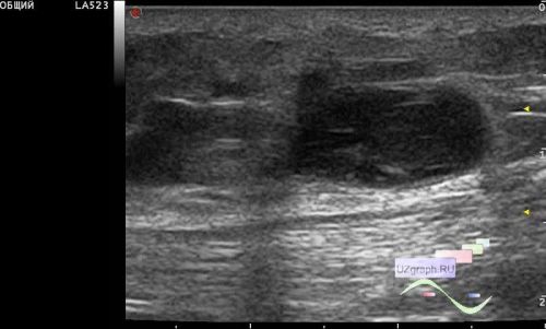
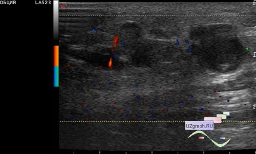
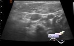
On the current follow-up ultrasound, in general, the echo picture described by another doctor in the protocol earlier is preserved, with the only difference that the largest of the lesions at the current follow-up ultrasound has a slightly smaller size: in the lower third of the anterior surface of the right thigh in the projection of the subcutaneous fatty tissue in front of the tendon of the quadriceps a group of an / hypoechoic lesions of oval and irregular curved shapes of various sizes (more than 5 pcs.), sometimes with an unclear margins: the largest 12 x 7 x 12 mm (differential diagnosis: lymphadenopathy, hematoma, etc.), the rest of the lesions are smaller, some of the smaller lesions have hyperechoic linear inclusions / septums in the structure, some visually communicate with each other (differential diagnosis: lymphangioma, lymphangiectasia, lymphostasis, venostasis, etc.). On the EDC / CDC, an increase in blood flow along the contour of some lesions and in the surrounding tissues is mapped.