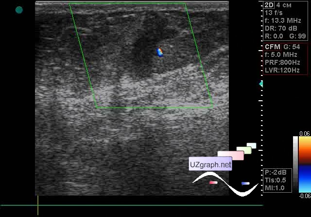
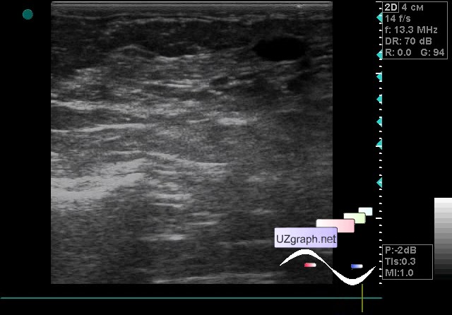
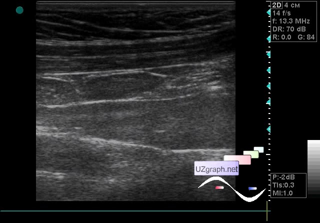
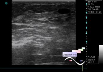
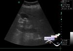
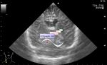
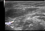
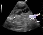
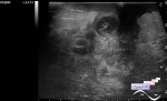
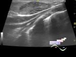
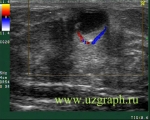
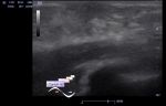
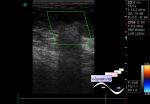
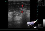
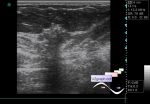
A 65-year-old patient came to an abdomen ultrasound because of the preparation program for the surgery, according to her words, an surgery in gynecology. In the process of the abdomen ultrasound, it turned out that recently the patient also underwent a mammogram, where by her words was found something and it seems that even this something in the gland is palpated. To her was also recommended to undergo breast ultrasound. Already after the abdomen ultrasound, I asked for the conclusion of the mammography, read it. In mammography conclusion describes the lesion of an irregular shape, with radiant contour, with a density of increased intensity, without disturbing the architectonics of the tissue, size of upto 1.5cm. Also on the same side, axillary LNs with a "susp. Mts" structure are described. But then at the bottom of the conclusion Birads 0 is indicated (additional examination is required). Recommendations are given for an examination by a mammologist, an ultrasound scan and a morphological verification of the lesion of Birads 0. Before reading this I recommended that the patient sign up for an ultrasound scan so as not to shift the schedule but after this reading I told her to lie down again. So let her go, and there ... On ultrasound in the projection of complaints on a dense lesion, a hypoechoic lesion of vertical orientation, of an irregular shape with a fuzzy contour is visualized; at CFM single signal is near, at PD single signal in the projection of lesion, size up to 1.5x0.7 cm (Birads 4-5, fig. 1-3 and video); beside the cyst up to 8x3mm(fig. 4 and video), on the CFM without blood flow. In the other mammary gland in the area of jumping of a probe (there would not be this probe jump - I would not have noticed this lesion) an isoechoic lesion of horizontal orientation is visualized, on the CFM without blood flow, up to 2x0.5 cm (fibroadenoma? Other? - fig. 5 and video). Subclavial LNs are enlarged on both sides to 1.5 cm (Fig. 6 and video). Urgent consultation of the mammologist was recommended with susp.cr. external link |