Log in    RU  


Main page :: Forum :: Ultrasound cases - Sonograms, cine-loops etc. ::

Birads 4-5


Tags: Breast sonography, Soft tissues sonography, GE Vivid 3, Images, Video, Clinical report, Adult

   Posts
 
  Birads 4-5
#1

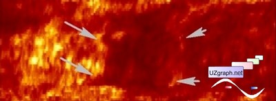
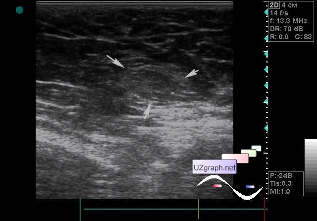
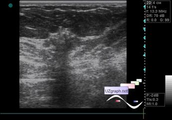
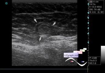
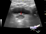
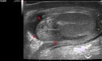
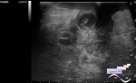
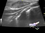
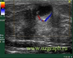
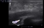
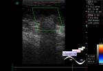
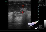
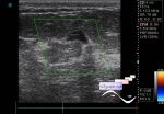
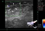
    A 57-year-old patient came for an ultrasound screening of the mammary glands; previously nothing was detected.

    In the left mammary gland at 5 o' clock a zone of medium echogenicity with a fuzzy uneven contour and a pronounced acoustic shadow and punctate echogenic foci (presumably microcalcifications), vertical orientation, up to 9 mm in size is visualized(birads 4-5?).

    In the left axillary region, 2 LNs of an atypical echo pattern (replacement by fat? mts?) up to 11x8mm, 12x9mm (attached files 4-5).

    In the right subclavian area single LN up to 9x5mm (attached file 6).

    Urgent consultation of mammologist and mammography was recommended.

    Ps. Attachment 3 - coronary view of birads 4-5 lesion.

    external link

:: attachments(7) ::

:: file 1 ::

:: file 2 ::

:: file 3 ::

:: file 4 ::

:: file 5 ::

:: file 6 ::

:: file 7 ::


 Random 
Ultrasound cases - Pain in the right iliac region
    

Ultrasound cases - Ovarian apoplexy
    

Ultrasound cases - Penetrating wound of the submandibular region
    

Ultrasound cases - Testicular torsion after surgery and thyroid
    

Ultrasound cases - Gallbladder duplication & bright kidneys
    

 Dictionary 
Freeze

Aliasing doppler artifact

BSA

CFM

SV

PSV

M-mode

EFW

LAP

HR


 Calculators 
Body surface area

Teicholz systolic cardiac function

Resistive index

Qp:Qs

Myocardial mass


 Similar topics 
Ultrasound cases - Mastitis and cysts
    

Ultrasound cases - Neonatal mastitis
    

Ultrasound cases - Adolescent Gynecomastia
    

Ultrasound cases - Subacute mastitis (cyst with inflammation) in a teenager
    

Ultrasound cases - Breast Cysts
    

Ultrasound cases - Infant isolated thelarche
    

Ultrasound cases - Another Birads 4-5
    

Ultrasound cases - Star-like lesion
    

Ultrasound cases - Breast lesions 3
    

Ultrasound cases - Milky ducts