Log in    RU  


Main page :: Forum :: Ultrasound cases - Sonograms, cine-loops etc. ::

Periventricular hyperechogenicity


Tags: Brain sonography(Neurosonography), GE Logiq 400 MD, Clinical report, Pediatric

   Posts
 
  Periventricular hyperechogenicity
#1

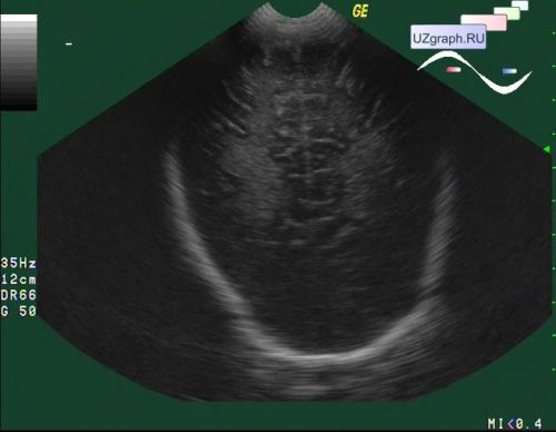
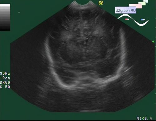
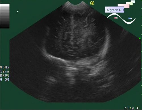
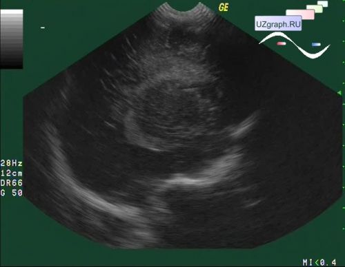
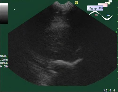
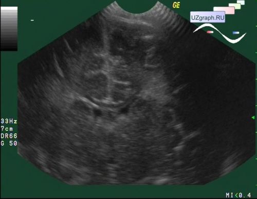
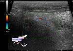
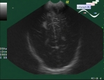
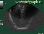
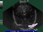
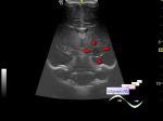
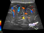
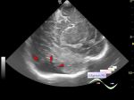
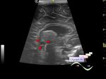
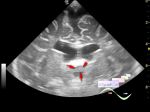
    An infant 1 month old came in a public clinic for screening.

    "Periventricular hyperechogenicity is a normal variant when it is smooth and less echo-rich on ultrasound than the choroid plexus. It is due to parenchymal immaturity." - The WHO Manual of diagnostic ultrasound.


 Random 
On-call room - And what is your gel, sterile?

Ultrasound cases - Gluteal area oedema / panniculitis
    

On-call room - Orange worm in medical equipment

Ultrasound cases - The lesion of the left thalamus
    

Ultrasound cases - Supposed renal hemangioma (angiomyolipoma)
    

 Ad 


 Dictionary 
LVLs

TI-RADS

Aliasing doppler artifact

LAP

ESV

GS

Freeze

LVLd

HR

ESD


 Calculators 
Measuring distance, area, circumference and angle on an image

Myocardial mass

Pulmonary artery pressure

Teicholz formula ejection fraction

Gestational age by Ultrasound


 Similar topics 
Ultrasound cases - Echogenic brain foci
    

Ultrasound cases - Subdural hemorrhage 2
    

Ultrasound cases - Willis circle at ultrasound
    

Ultrasound cases - Combined (internal and external) hydrocephalus
    

Ultrasound cases - Subdural hemorrhage
    

Ultrasound cases - Cyst in the projection near the posterior horn of the lateral ventricle
    

Ultrasound cases - Subdural collection
    

Ultrasound cases - Anechogenic inclusion in the posterior cranial fossa
    

Ultrasound cases - Cyst of the base of the brain
    

Ultrasound cases - Interthalamic adhesion aberration