Log in    RU  


Main page :: Forum :: Ultrasound cases - Sonograms, cine-loops etc. ::

Subdural collection


Tags: Brain sonography(Neurosonography), GE Logiq E, Clinical report, Pediatric

   Posts
 
  Subdural collection
#1

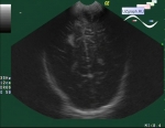
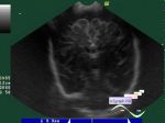
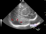
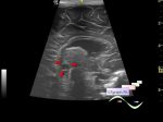
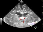
    A 5-month-old child came for a follow-up ultrasound due to increased periventricular echogenicity (immaturity of the brain); the craniocerebral space examined by ultrasound at the age of 1 month was normal.

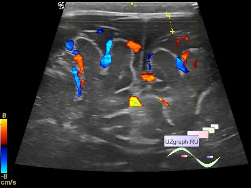
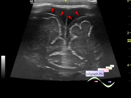
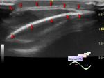
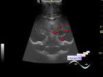
    On the current ultrasound, subconvexital (craniocerebral) space up to 9 mm (normal < 6mm): subdural space up to 5 mm (differential diagnosis: subdural hematoma/ hygroma).


 Random 
Ultrasound cases - Ladd's syndrome after surgery
    

Ultrasound cases - Star-like lesion
    

Ultrasound cases - Thyroid lesions 2
    

Ultrasound cases - Good vs Evil - thyroid 4
    

Ultrasound cases - Lesion in the projection of the adrenal gland
    

 Dictionary 
TAV

VTI

ESD

PW

Gain

IVC

Color M-mode

Freeze

PSV

SV


 Calculators 
Pressure gradient

Gestational age by Ultrasound

Myocardial mass

Estimated fetal weight by Ultrasound

Resistive index


 Similar topics 
Ultrasound cases - Echogenic brain foci
    

Ultrasound cases - Subdural hemorrhage 2
    

Ultrasound cases - Willis circle at ultrasound
    

Ultrasound cases - Periventricular hyperechogenicity
    

Ultrasound cases - Combined (internal and external) hydrocephalus
    

Ultrasound cases - Subdural hemorrhage
    

Ultrasound cases - Cyst in the projection near the posterior horn of the lateral ventricle
    

Ultrasound cases - Anechogenic inclusion in the posterior cranial fossa
    

Ultrasound cases - Cyst of the base of the brain
    

Ultrasound cases - Interthalamic adhesion aberration