An infant came to the ED of Children City Hospital with suspicion of intussusception, has an ultrasound scan at other health care facilities, according to the results of which intussusception was suspected, however, in the ultrasound scan paper that the parents brought with them, there was no mention of the target sign, if my memory serves me, there was described a part of the intestine with a thickened wall in the right iliac region with the following conclusion - a spasmodic bowel loop (intussusception?).
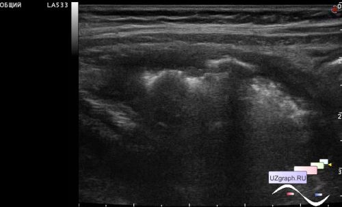
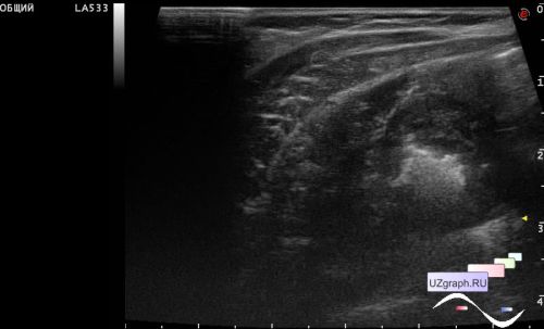
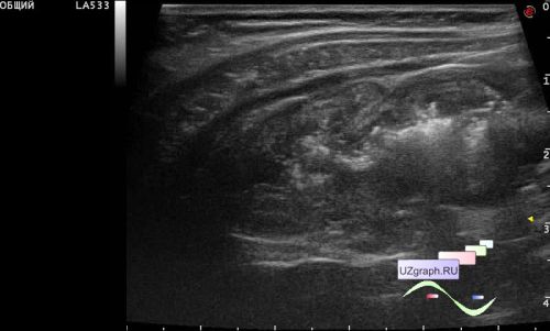
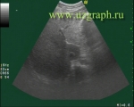
On ultrasound, a part of the intestine without peristalsis and with a thickened wall was visualized in the right ileal region, but it did not look like intussusception, but rather an inflammatory process (?), I described this picture as an HOLS (Hollow Organ Lesion Sign) type of the pseudokidney sign ... I personally explained everything verbally to the surgeon, who turned out to be unfamiliar with the HOLS (It's russian term which include target sign, pseudokidney sign, etc) abbreviation, including about the uncharacteristic nature of this ultrasound symptom (pseudokidney) for intussusceptions, as it later turned out that the abbreviation HOLS, like the expanded version of the term, is unknown not only to clinicians, but and ultrasound specialists.
Coming back to the case, despite my explanations, the surgeon still translate my record as a sign of intussusception! After the procedure of air expansion of the intussusception supposed by the clinicians, a follow-up ultrasound was prescribed, and everything remained the same on the ultrasound.
The next day, the child underwent another follow-up ultrasound scan by another doctor, where an opinion was expressed about the probable infectious process and the absence of signs of intussusception. Which was told me in a critical style, something like this - "there is no intussusception about which you wrote" - although I did not write about it, I wrote HOLS. It is good that the 4th volume of the 1997 edition of V.V. Mitkov's ultrasound manual accidentally turned out to be at hand, in which, to the surprise of the critically minded ultrasound specialists, HOLS was described as I told.
PS. Although in the end of video you can see an area of the intestine that can be called a mushroom sign, and it can correspond to both intussusception in the ileocecal region and ileotiflitis ...