Log in    RU  


Main page :: Forum :: Ultrasound cases - Sonograms, cine-loops etc. ::

Suspected retroperitoneal hematoma


Tags: Abdomen sonography, Urinary tract sonography, Images, Video, Clinical report, Esaote MyLab 70, Pediatric

   Posts
 
  Suspected retroperitoneal hematoma
#1

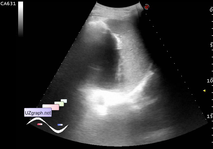
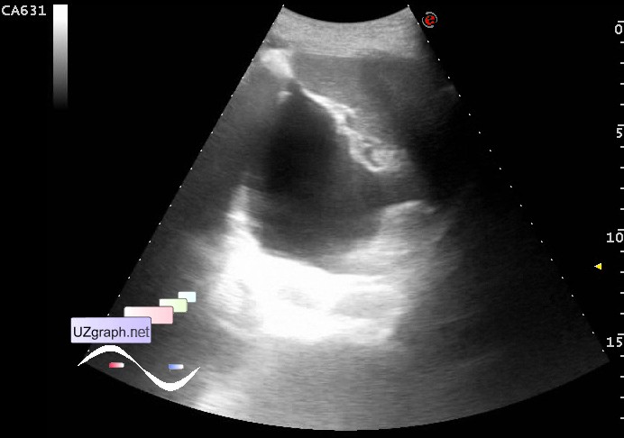
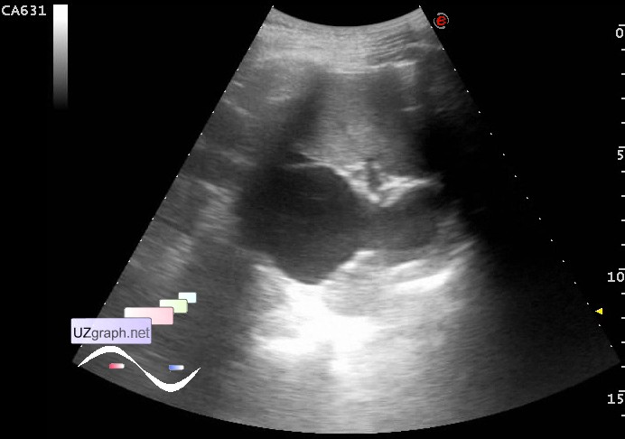
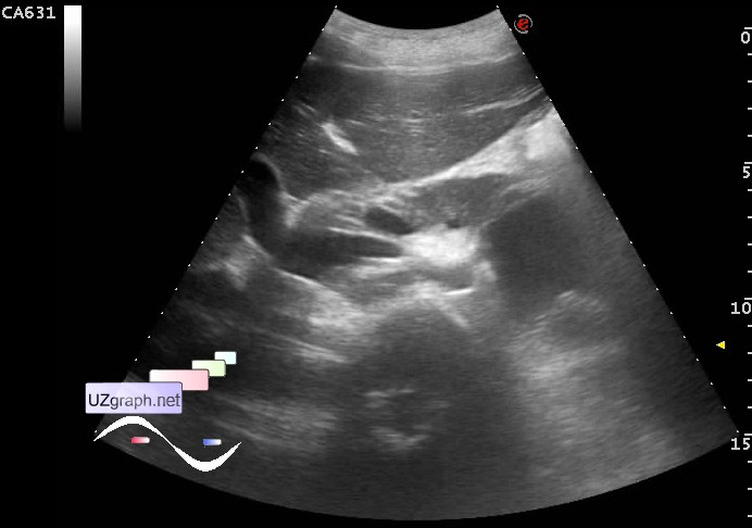
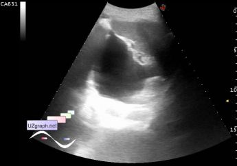
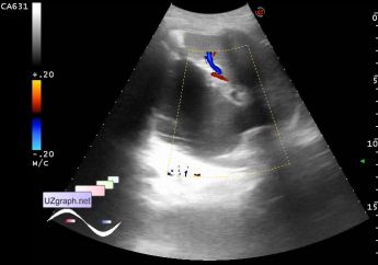
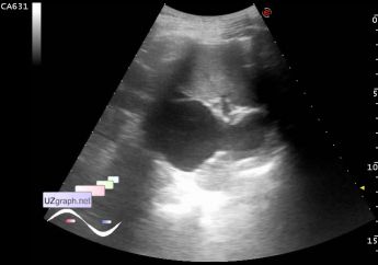
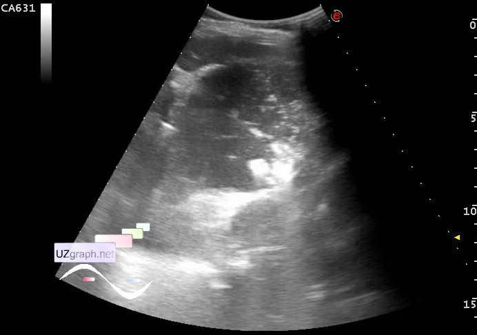
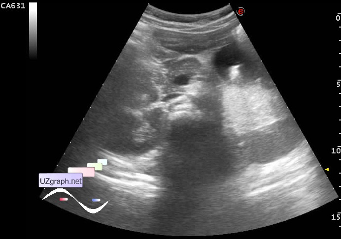
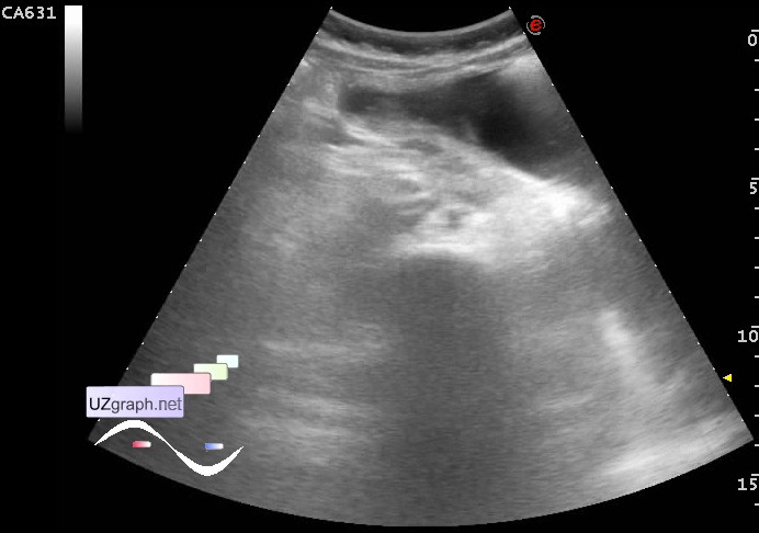
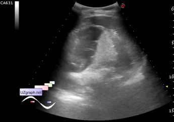
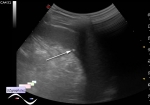
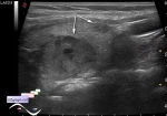
    A child of 14 years old with suspected damage of internal organs.

    According to relatives a week ago was hit by a car, crossing the road in the wrong place, as reported relatives, doctors, once they heard about it they immediately dragged the child to the work and then had a MRI ,CT & ultrasound. MRI have found a cyst, hematoma was labeled on CT, ultrasound also found the hematoma. CT sent them to the US because this institution partly traumatological and sonologist of course the traumatologist, but such founding was not the profile of this institution, so they were sent them to us, and our surgeons of course sent them to our US, although were all photocopies ...

    In the current US all exactly as it was described on the previous ultrasound, CT, MRI earlier ...

    In the projection posterior to tail of the pancreas, anterior to the left kidney, medial to the spleen there is a mainly anehoic irregular shape lesion, up to approximately 9 cm, at CFM without bloodflow (hematoma?)

    And as well as fellow diagnosticians I also verbally expressed doubts that it could be something else, and that the hematoma is suspected only on the basis of history of the trauma.

    external link

:: attachments(10) ::

:: file 1 ::

:: file 2 ::

:: file 3 ::

:: file 4 ::

:: file 5 ::

:: file 6 ::

:: file 7 ::

:: file 8 ::

:: file 9 ::

:: file 10 ::

  03:54 15-04-2016
  just full stomach
#2

    PS. I remembered the case, about which we were told 10 years ago, it's about the stomach, which is a few hours don't take a rest for sonologists. Therefore, there is another video clip with full stomach to compare - are they matched?

    A child of 4 years old with suspected appendicitis, with a full stomach ...

:: attachments(6) ::

:: file 1 ::

:: file 2 ::

:: file 3 ::

:: file 4 ::

:: file 5 ::

:: file 6 ::


 Random 
Ultrasound cases - Recurrent shin trauma
    

Ultrasound cases - Clinically suspected appendicitis
    

Ultrasound cases - Star-like lesion
    

Ultrasound cases - Rested
    

Ultrasound cases - Hemorrhagic ovarian cyst
    

 Dictionary 
BSA

LAP

PD

Ved

LN

Color M-mode

PASP

M-mode

Gain

GS


 Calculators 
Volume (ellipse formula)

Myocardial mass

Gestational age by Ultrasound

Resistive index

TI-RADS


 Similar topics 
Ultrasound cases - Hirschsprung disease on ultrasound
    

Ultrasound cases - Intestinal Infiltrate
    

Ultrasound cases - Abdominal cryptorchidism
    

Ultrasound cases - Diffuse heterogeneous liver
    

Ultrasound cases - Postoperative abdominal wall fistula
    

Ultrasound cases - Stump appendicitis
    

Ultrasound cases - Liver scar after resection
    

Ultrasound cases - Subdiaphragmatic movable fluid collection (UFO)
    

Ultrasound cases - Lesion of the spleen or the case when it is easy to not notice
    

Ultrasound cases - I’m normal but others ...