Log in    RU  


Main page :: Forum :: Ultrasound cases - Sonograms, cine-loops etc. ::

Venous lacunae


Tags: Brain sonography(Neurosonography), Medison Sonoace R7, Images, Video, Clinical report, Pediatric

   Posts
 
  Venous lacunae
#1

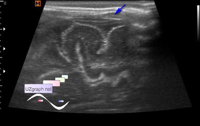
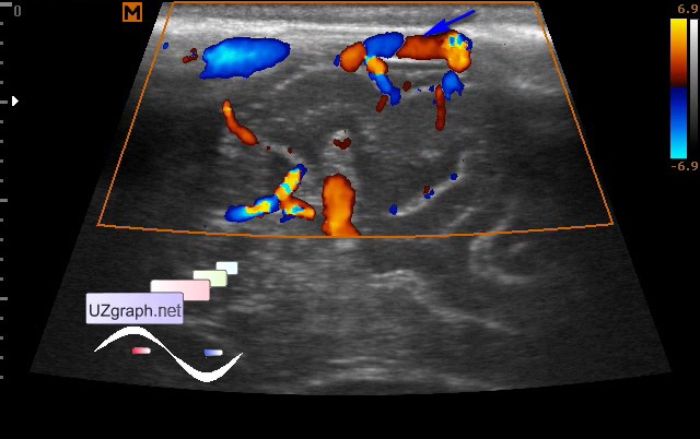
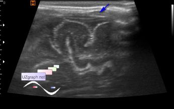
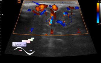
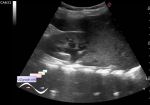
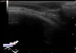
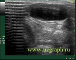
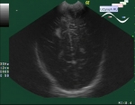
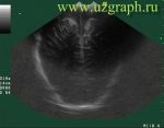
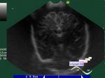
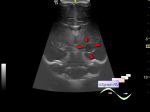
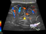
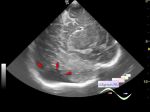
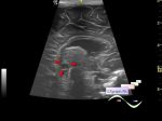
    Child 8 months. With suspicion of intracranial hypertension due to the results of brain ultrasound in the private medical center, the protocol of the previous brain ultrasound they didn't bring (apparently decided to check me or them).

    I looked at what the commercial medicare could find fault with.

    On the ultrasound subconvexitally next to the superior sagittal sinus pulsating anechoic structures of the oval / tubular shape are visualized, more at the left, with a thickness up to 2mm, with blood flow at CFM (venous lacunae), Vmax up to 5 cm / sec, the shape of the spectrum by the venous type, flowing into the superior sagittal sinus, Vmax in Galen vein 11cm / s.

    lateral / venous lacunae - on the attached files are indicated by a blue arrow, the white arrow indicates the superior sagittal sinus.

    Additionally:

    external link

    external link

    external link

:: attachments(5) ::

:: file 1 ::

:: file 2 ::

:: file 3 ::

:: file 4 ::

:: file 5 ::


 Random 
Ultrasound cases - Perinephric lesion
    

Ultrasound cases - Thickening of the spermatic cord in a teenager
    

Ultrasound cases - Diffuse heterogeneous liver
    

Ultrasound cases - Cystic-solid volume of the thyroid gland
    

Ultrasound cases - Record size appendicitis
    

 Dictionary 
B-mode

MSD

LVESD

LN

CO

LVEDV

Gain

BPD

TVUS

IVSd


 Calculators 
Pulmonary artery pressure

Resistive index

Myocardial mass

Gestational age by Ultrasound

Volume (ellipse formula)


 Similar topics 
Ultrasound cases - Echogenic brain foci
    

Ultrasound cases - Subdural hemorrhage 2
    

Ultrasound cases - Willis circle at ultrasound
    

Ultrasound cases - Periventricular hyperechogenicity
    

Ultrasound cases - Combined (internal and external) hydrocephalus
    

Ultrasound cases - Subdural hemorrhage
    

Ultrasound cases - Cyst in the projection near the posterior horn of the lateral ventricle
    

Ultrasound cases - Subdural collection
    

Ultrasound cases - Anechogenic inclusion in the posterior cranial fossa
    

Ultrasound cases - Cyst of the base of the brain