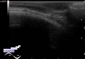
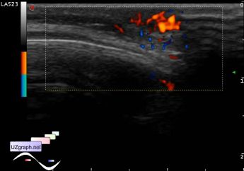
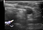
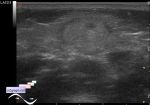
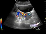
Hemangioma in the back of the head
Tags: Soft tissues sonography, Images, Video, Clinical report, Esaote MyLab 70, Pediatric
| Posts | |||
| Hemangioma in the back of the head | #1 |
| |||||
:: file 1 ::
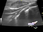
 :: file 2 ::
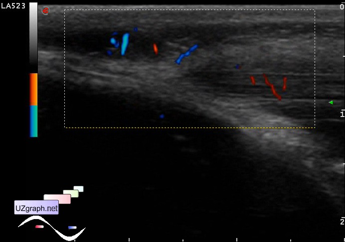
 :: file 3 ::
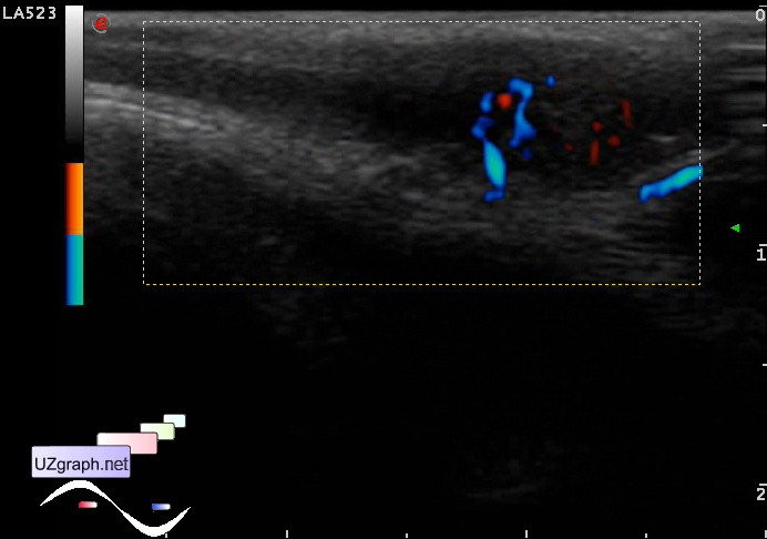
 :: file 4 ::
 :: file 5 ::
 :: file 6 :: | |||||