Log in    RU  


Main page :: Forum :: Ultrasound cases - Sonograms, cine-loops etc. ::

Lateral neck cyst/ branchial cleft cyst


Tags: Soft tissues sonography, GE Logiq 400 MD, Clinical report, Pediatric

   Posts
 
  Lateral neck cyst/ branchial cleft ...
#1

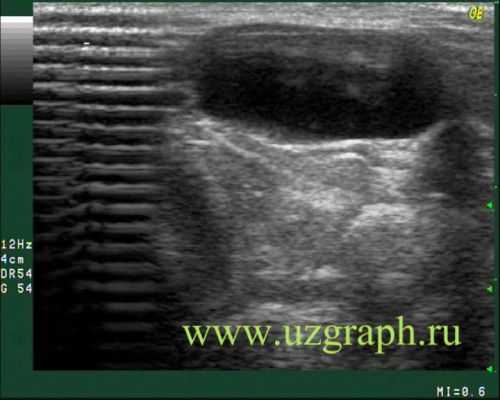
    Parents of the 5-year-old child turned to the clinic with complaints that a few days ago a painful lesion appeared on the anterior-lateral surface of the child neck, accompanied by an increase in body temperature, which was initially regarded as a lymph node, but after consulting a surgeon, the child was referred to Ultrasound of the neck to exclude other pathology. By the time of the study, the mass lesion had decreased in size, painless. On ultrasound in the projection of complaints, an anechoic oval lesion without blood flow at CFM is visualized (most likely a lateral cyst of the neck/ branchial cleft cyst ).


 Random 
Ultrasound cases - Kidney blockage
    

Ultrasound cases - Idiopathic low-grade fever?
    

Ultrasound cases - Neonatal hemorrhagic syndrome
    

Ultrasound cases - Supravalvular aortic stenosis
    

Ultrasound cases - Rectus abdominis muscle contusion
    

 Dictionary 
PSV

GA

EDD

PD

PA

Doppler angle

LN

FNA

MPAP

LVESV


 Calculators 
Type of an infant hip joint

Pressure gradient

Estimated fetal weight by Ultrasound

Thyroid volume

Teicholz systolic cardiac function


 Similar topics 
Ultrasound cases - Penetrating injury of the femur
    

Ultrasound cases - Cephalohematoma
    

Ultrasound cases - Urachus fistula
    

Ultrasound cases - Cranial mass
    

Ultrasound cases - Pilonidal coccyx cyst
    

Ultrasound cases - Palm fistula, in search of pencil lead
    

Ultrasound cases - Postoperative abdominal wall fistula
    

Ultrasound cases - Nucks cyst
    

Ultrasound cases - Paraproctitis
    

Ultrasound cases - Submandibular area lesion