Log in    RU  


Main page :: Forum :: Ultrasound cases - Sonograms, cine-loops etc. ::

Ulnar area shoulder mass


Tags: Soft tissues sonography, Images, Video, Clinical report, Esaote MyLab 70, Pediatric

   Posts
 
  Ulnar area shoulder mass
#1

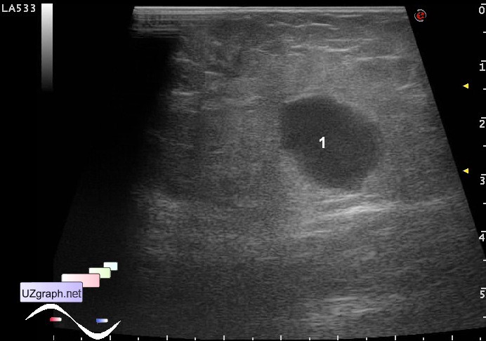
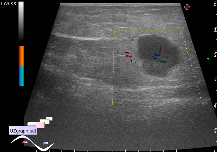
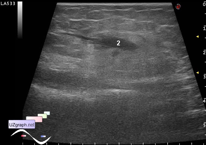
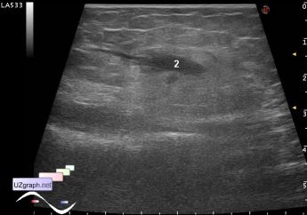
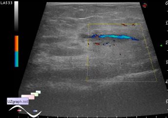
    Teen girl with suspected mass in the distal third of the medial surface of the right shoulder is directed to the ultrasound.

    At ultrasound at a depth of about 1.5 cm there is a hypoechoic lesion about 1.5 cm in diameter, with blood flow at DPD(lymph node? marked as 1 at image). Near this lesion visualized a tubular structure looks like a vein thrombosis (recanalization? marked as 2 at image).

    external link

:: attachments(5) ::

:: file 1 ::

:: file 2 ::

:: file 3 ::

:: file 4 ::

:: file 5 ::


 Random 
Ultrasound cases - Lesion in the female pelvis
    

Ultrasound cases - Calcific abdominal lymphadenopathy
    

Ultrasound cases - Thigh lesions
    

Ultrasound cases - Lesion in pancreas head
    

Ultrasound cases - Shunt and cyst of brain ventricle
    

 Dictionary 
CO

HR

SV

RAP

LN

TVUS

PRF

LVEDV

GA

LVOT


 Calculators 
Type of an infant hip joint

Pulmonary artery pressure

TI-RADS

Qp:Qs

Resistive index


 Similar topics 
Ultrasound cases - Skin shell
    

Ultrasound cases - Skin lesion in the projection of the surgical suture
    

Ultrasound cases - Purulent / Suppurative lymphadenitis
    

Ultrasound cases - Hematoma of the gluteal region
    

Ultrasound cases - Thickening and heterogeneity of the intercostal space soft tissues
    

Ultrasound cases - Chest wall lesion
    

Ultrasound cases - Hockey Elbow
    

Ultrasound cases - Combination of left shoulder lesions
    

Ultrasound cases - Infiltrate/ phlegmon of the wrist
    

Ultrasound cases - Subcutaneous lesion above the sternum