Log in    RU  


Main page :: Forum :: Ultrasound cases - Sonograms, cine-loops etc. ::

LSVC and PDA


Tags: Cardiac sonography(Echocardiography), Esaote MyLab 70, Images, Video, Clinical report, Pediatric

   Posts
 
  LSVC and PDA
#1

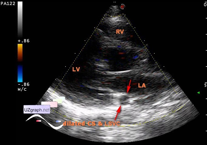
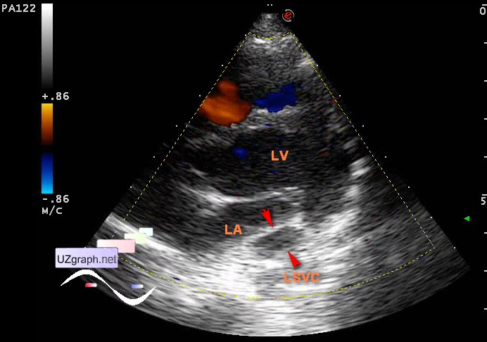
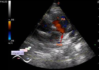
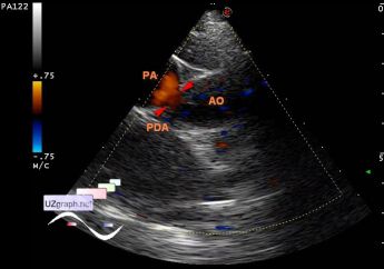
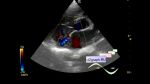
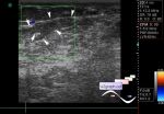
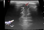
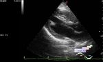
    Child 6 years-old came for control ultrasound of the heart, in the history - PDA(patent ductus arteriosus) which is closed by the data of the previous ultrasound and LSVC (left superior vena cava).

    At the current US is visualized LSVC draining into dilated coronary sinus (CS) and PDA in typical places(at the base of the posterior leaflet of mitral valve(dilated CS) and at the pulmonary artery bifurcations, respectively).

    external link

:: attachments(5) ::

:: file 1 ::

:: file 2 ::

:: file 3 ::

:: file 4 ::

:: file 5 ::


 Random 
Ultrasound cases - Acute appendicitis, typhlitis
    

Ultrasound cases - "Tricky" IVSD
    

Ultrasound cases - Lesions of mammary glands 2
    

Ultrasound cases - 99 Luftballons
    

Ultrasound cases - Alien
    

 Dictionary 
Color M-mode

LVESD

Gain

LN

Doppler angle

PG

MPAP

RVOT

AC

RAP


 Calculators 
Body surface area

Estimated fetal weight by Ultrasound

Myocardial mass

Volume (ellipse formula)

Teicholz systolic cardiac function


 Similar topics 
Ultrasound cases - Bicuspid aortic valve 2
    

Ultrasound cases - Mitral valve cyst
    

Ultrasound cases - Electrode in the right heart after installing of heart pacemaker
    

Ultrasound cases - Chylopericardium, lung atelectasis, cardiomegaly, ascites
    

Ultrasound cases - Asymmetric LVH, aortic regurgitation
    

Ultrasound cases - Coronary fistula from LCA?
    

Ultrasound cases - Floating IAS aneurysm
    

Ultrasound cases - Aortic Stenosis
    

Ultrasound cases - Floating Accessory Pulmonary Valve Tissue
    

Ultrasound cases - Hyperechoic linear structure in the left atrium