Log in    RU  


Main page :: Forum :: Ultrasound cases - Sonograms, cine-loops etc. ::

Atypical axillary LNs


Tags: Soft tissues sonography, GE Vivid 3, Images, Video, Clinical report, Adult

   Posts
 
  Atypical axillary LNs
#1

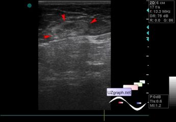
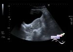
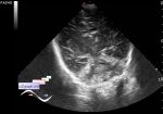
    The patient 66 years old female came to the screening ultrasound of the breast, no additional lesions were found in the mammary glands.

    But single axillary LNs(lymph nodes) of an atypical structure (mainly hyperechoic with an unclear contour - replacement by fat? Other?) were found on both sides, up to 2 cm in length, on the CFM on the one side with blood flow.

    PS. Some people think that replacement LNs by fat is practically always the age norm, but -

    " abundant replacement of the lymph node with fat is highly unusual and may appear as metastatic lymph node disease in the course of fat-predominant liposarcomas or in the case of coeliac disease complicated by cavitating lymph node syndrome. In this case report, a patient with chronic lymphocytic leukaemia/small lymphocytic lymphoma who demonstrated an increasing abundance of macroscopic fat in the diseased lymph nodes is presented."

    external link

:: attachments(5) ::

:: file 1 ::

:: file 2 ::

:: file 3 ::

:: file 4 ::

:: file 5 ::


 Random 
Ultrasound cases - Fenestration of AS / Multiple ASD
    

Ultrasound cases - Scrotum pearls
    

Ultrasound cases - Nephrocalcinosis
    

Ultrasound cases - Gallbladder polyp simulation
    

Ultrasound cases - Cyst in the projection near the posterior horn of the lateral ventricle
    

 Dictionary 
HR

EF

LN

Color M-mode

EDV

CO

TI-RADS

EFW

PADP

PASP


 Calculators 
Resistive index

Pressure gradient

Gestational age by Ultrasound

Teicholz systolic cardiac function

Pulmonary artery pressure


 Similar topics 
Ultrasound cases - Penetrating injury of the femur
    

Ultrasound cases - Cephalohematoma
    

Ultrasound cases - Urachus fistula
    

Ultrasound cases - Cranial mass
    

Ultrasound cases - Pilonidal coccyx cyst
    

Ultrasound cases - Palm fistula, in search of pencil lead
    

Ultrasound cases - Postoperative abdominal wall fistula
    

Ultrasound cases - Nucks cyst
    

Ultrasound cases - Paraproctitis
    

Ultrasound cases - Submandibular area lesion