Log in    RU  


Main page :: Forum :: Ultrasound cases - Sonograms, cine-loops etc. ::

Ileostoma stenosis and nephropathy


Tags: Abdomen sonography, Urinary tract sonography, Gastrointestinal sonography, Esaote MyLab 70, Clinical report, Pediatric

   Posts
 
  Ileostoma stenosis and nephropathy
#1

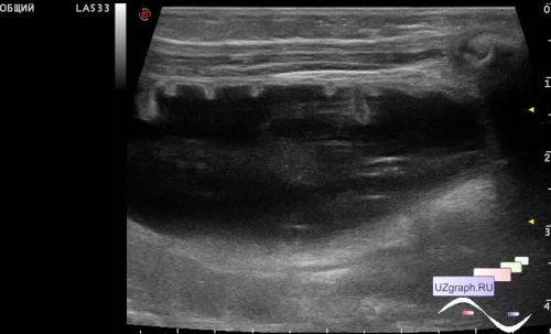
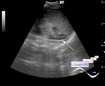
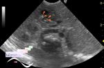
    An infant in the Children's City Clinical Hospital with ileostomy stenosis, is referred for an abdominal ultrasound.

    In the left abdomen, longitudinally to the body axis, there is an dilated intestine with fluid contents (stoma), the thickness of the rectal wall is up to 5 mm.

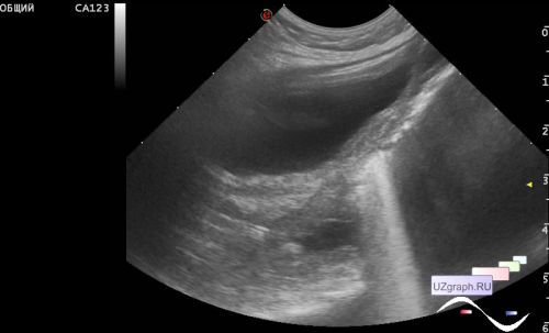
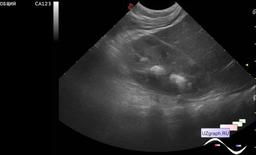
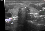
    In one of the kidneys in longitudinal views there are hyperechoic lesions along the pyramids (hyperechoic pyramids sign).


 Random 
Ultrasound cases - Curved small popliteal cyst
    

Ultrasound cases - Detorsion of the hydatide torsion
    

Ultrasound cases - Scrotum hydrocele
    

Ultrasound cases - Hyperechoic fat like inclusions in the scrotum
    

Ultrasound cases - Anechogenic inclusion in the posterior cranial fossa
    

 Dictionary 
DPD

B-mode

TV

TAV

EDD

LVPWd

PG

MSD

LMP

TVUS


 Calculators 
Myocardial mass

Volume (ellipse formula)

Body surface area

Pressure gradient

Thyroid volume


 Similar topics 
Ultrasound cases - Diffuse heterogeneous liver
    

Ultrasound cases - Postoperative abdominal wall fistula
    

Ultrasound cases - Stump appendicitis
    

Ultrasound cases - Liver scar after resection
    

Ultrasound cases - Subdiaphragmatic movable fluid collection (UFO)
    

Ultrasound cases - Lesion of the spleen or the case when it is easy to not notice
    

Ultrasound cases - I’m normal but others ...
    

Ultrasound cases - Single doubled kidney and Mullerian duct cyst in the infant
    

Ultrasound cases - Condition after removal of the spleen cyst
    

Ultrasound cases - Portal vein air in the newborn