Log in    RU  


Main page :: Forum :: Ultrasound cases - Sonograms, cine-loops etc. ::

The lesion of the left thalamus


Tags: Brain sonography(Neurosonography), Gastrointestinal sonography, Images, Video, Clinical report, Esaote MyLab 70, Pediatric

   Posts
 
  The lesion of the left thalamus
#1

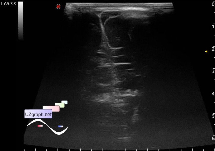
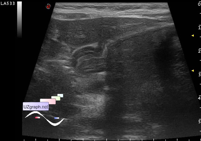
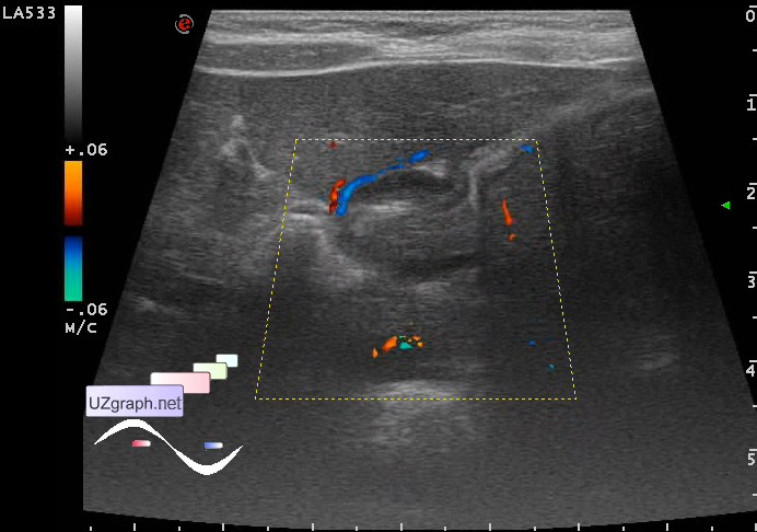
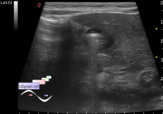
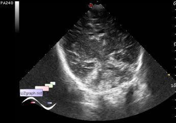
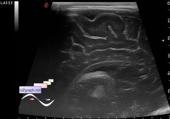
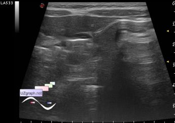
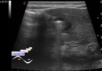
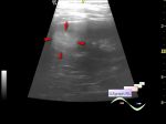
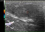
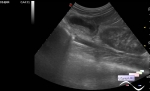
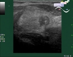
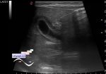
    Baby 2 month ultrasound screening at the age of 1 month, including neurosonography(NSG) - without a pathology.

    On the same day he was sent to one of child hospital(CH) in Moscow because of the prolonged jaundice, where on the next day was done NSG - in the projection of basal ganglia (thalamus) in the left is determined the triangular shape focus of increased echogenicity with indistinct contours 11x7mm, with no signs of increased vascular pattern at CFM, the exact morphological picture at the time of exam is difficult to determine. Also in this CH suspected the cytomegalovirus infection, pilorospazm / developing pyloric stenosis (the wall thickness of the pyloric stomach 2.5-3mm), increased echogenicity of the kidneys.

    Recommendations repeat NSG and abdominal US, kidney US after 1 week (in the amplification of regurgitation before).

    At the current ultrasound in the left thalamus visualized two intimately located hyperechoic structure, one linear 8x3mm (talamostriarnaya vasculopathy?) and second round shape 5x8x6mm (?).

    external link

    Stomach with heteroechoic content, pyloric portion - closed: of 14 mm in length, diameter 10.6 mm, muscular wall to 3.5 mm; open: to 2.5 mm muscular wall, pyloric portion opens, visualised the food passage (pilorospazm).

    external link

    Kidneys are normal.

    Recommended CT / MRI of the brain, consulting neurosurgeon / neurologist.

:: attachments(10) ::

:: file 1 ::

:: file 2 ::

:: file 3 ::

:: file 4 ::

:: file 5 ::

:: file 6 ::

:: file 7 ::

:: file 8 ::

:: file 9 ::

:: file 10 ::

  19:04 09-11-2017
 
#2

    Today was at the lecture about strokes in infants, in particular, there was presented a case of a stroke on ultrasound - the initial brain ultrasound without changes and repeated ultrasound after 2 hours with a large zone (several cm) of hyperechogenicity.

    So 2 hours is enough.

  02:43 12-04-2022
 
#3

    "Risk factors for pediatric stroke."

    See table 1 at external link


 Random 
Ultrasound cases - Cyst near UB
    

Ultrasound cases - Chest wound with barbed wire
    

Ultrasound cases - Lymph node purulent destruction
    

Ultrasound cases - Milky ducts
    

Ultrasound cases - Diaphragmatic hernia, condition after surgery
    

 Dictionary 
IVSd

LVOT

ESV

PADP

AC

TV

PW

Qp-Qs

GA

B-mode


 Calculators 
Volume (ellipse formula)

Type of an infant hip joint

Pressure gradient

Body surface area

Myocardial mass


 Similar topics 
Ultrasound cases - Echogenic brain foci
    

Ultrasound cases - Subdural hemorrhage 2
    

Ultrasound cases - Willis circle at ultrasound
    

Ultrasound cases - Periventricular hyperechogenicity
    

Ultrasound cases - Combined (internal and external) hydrocephalus
    

Ultrasound cases - Subdural hemorrhage
    

Ultrasound cases - Cyst in the projection near the posterior horn of the lateral ventricle
    

Ultrasound cases - Subdural collection
    

Ultrasound cases - Anechogenic inclusion in the posterior cranial fossa
    

Ultrasound cases - Cyst of the base of the brain