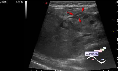
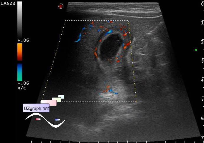
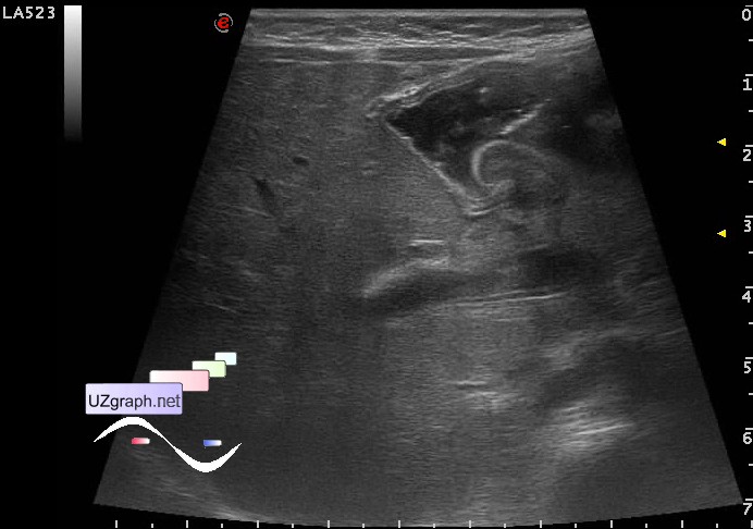
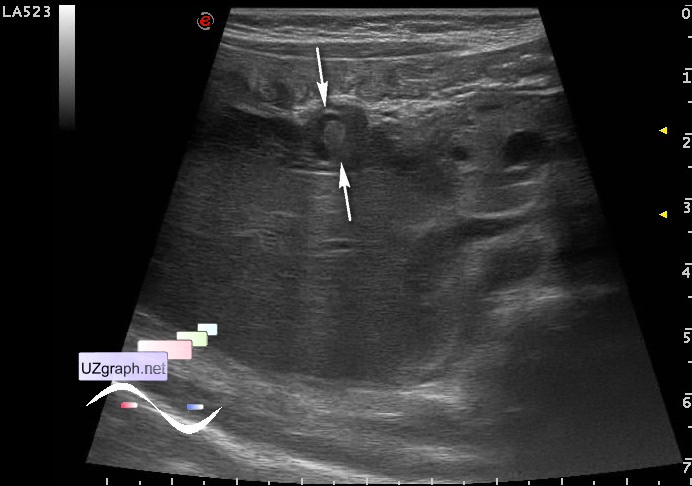
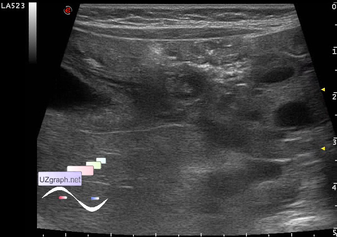
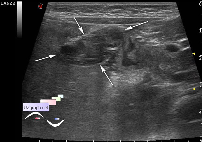
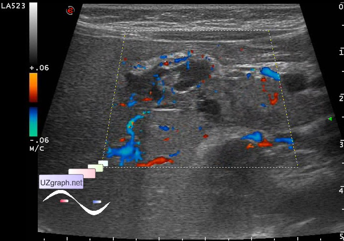
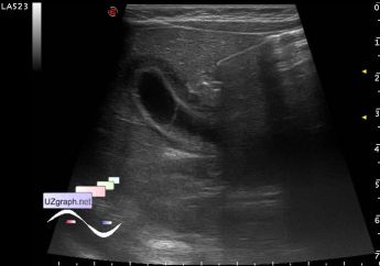
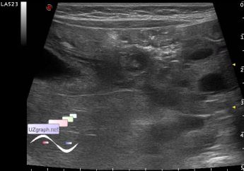
Intestinal obstruction, GB wall thickening
Tags: Abdomen sonography, Gastrointestinal sonography, Images, Video, Clinical report, Esaote MyLab 70, Pediatric
| Posts | |||
| Intestinal obstruction, GB wall thi... | #1 |
| |||||
:: file 1 ::
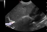
 :: file 2 ::
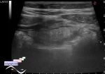
 :: file 3 ::
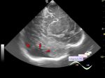
 :: file 4 ::
 :: file 5 ::
 :: file 6 ::
 :: file 7 ::
 :: file 8 :: | |||||


| 06:36 05-10-2019 | #2 |
| |||||
| 17:06 28-04-2024 | #3 |
| |||||