Log in    RU  


Main page :: Forum :: Ultrasound cases - Sonograms, cine-loops etc. ::

Milky ducts


Tags: Breast sonography, GE Vivid 3, Images, Video, Clinical report, Adult

   Posts
 
  Milky ducts
#1

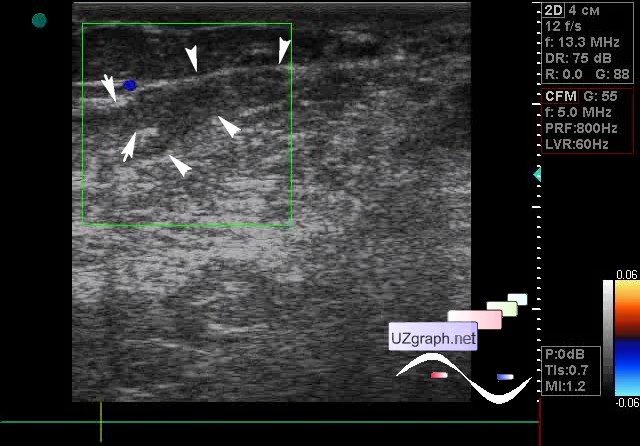
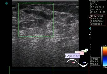
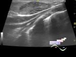
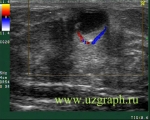
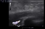
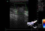
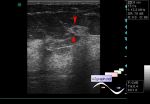
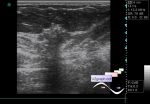
    A 79-year-old patient came to a screening ultrasound examination of the mammary glands; recently there was an injury, including the left mammary gland.

    At ultrasonography in the left mammary gland visualizes several ducts in the periphery region with hyperechoic contents type of sludge, without blood flow in the CFM, up to 3 mm in diameter (mastitis? Etc. - intraductal lesion?)

    For example,

    Intraductal papilloma

    external link

    external link

:: attachments(3) ::

:: file 1 ::

:: file 2 ::

:: file 3 ::


 Random 
Ultrasound cases - Hirschsprung's
disease after surgery


Ultrasound cases - Simple colloid nodule
    

Ultrasound cases - Intestine contusion
    

Ultrasound cases - Record size appendicitis
    

Ultrasound cases - Hydrosalpinx (saktosalpinx)
    

 Dictionary 
Doppler angle

Qp-Qs

HC

MC

PG

MSD

BPD

SV

M-mode

TAV


 Calculators 
Myocardial mass

Estimated fetal weight by Ultrasound

Pressure gradient

Resistive index

Pulmonary artery pressure


 Similar topics 
Ultrasound cases - Mastitis and cysts
    

Ultrasound cases - Neonatal mastitis
    

Ultrasound cases - Adolescent Gynecomastia
    

Ultrasound cases - Subacute mastitis (cyst with inflammation) in a teenager
    

Ultrasound cases - Breast Cysts
    

Ultrasound cases - Infant isolated thelarche
    

Ultrasound cases - Another Birads 4-5
    

Ultrasound cases - Star-like lesion
    

Ultrasound cases - Birads 4-5
    

Ultrasound cases - Breast lesions 3