Log in    RU  


Main page :: Forum :: Ultrasound cases - Sonograms, cine-loops etc. ::

Cholesterol pseudopolyp?


Tags: Abdomen sonography, GE Vivid 3, Images, Video, Clinical report, Adult

   Posts
 
  Cholesterol pseudopolyp?
#1

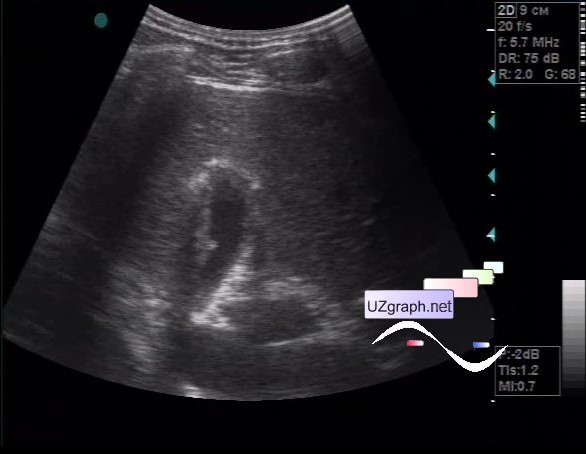
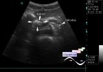
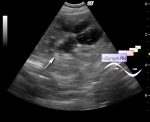
    The patient 23 years old, came for the control ultrasound of the gallbladder polyp.

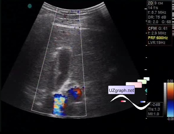
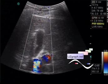
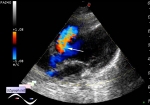
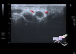
    On ultrasound in the projection of the lateral wall the parietal lesion up to 3 mm is visualized, the blood flow was not visualized at CFM (presumably cholesterol pseudopolyp).

    external link

:: attachments(3) ::

:: file 1 ::

:: file 2 ::

:: file 3 ::


 Random 
On-call room - How to become a sonographer

Ask DMS-professionals - Answering your questions

Ultrasound cases - Breast Cysts
    

Ultrasound cases - Spleen cyst
    

Ultrasound cases - Linea alba hernia vs lipoma part trois
    

 Dictionary 
LVEDV

LVESD

LVPWd

MSD

TAV

LMP

EF

IVSd

ESD

EFW


 Calculators 
Pulmonary artery pressure

Qp:Qs

Volume (ellipse formula)

Pressure gradient

Teicholz systolic cardiac function


 Similar topics 
Ultrasound cases - Hirschsprung disease on ultrasound
    

Ultrasound cases - Intestinal Infiltrate
    

Ultrasound cases - Abdominal cryptorchidism
    

Ultrasound cases - Diffuse heterogeneous liver
    

Ultrasound cases - Postoperative abdominal wall fistula
    

Ultrasound cases - Stump appendicitis
    

Ultrasound cases - Liver scar after resection
    

Ultrasound cases - Subdiaphragmatic movable fluid collection (UFO)
    

Ultrasound cases - Lesion of the spleen or the case when it is easy to not notice
    

Ultrasound cases - I’m normal but others ...