Log in    RU  


Main page :: Forum :: Ultrasound cases - Sonograms, cine-loops etc. ::

Lesion in pancreas head


Tags: Abdomen sonography, GE Vivid 3, Images, Video, Clinical report, Adult

   Posts
 
  Lesion in pancreas head
#1

    A 23-year-old female patient came to an abdomen ultrasound complaining of a change in the color of the stool.

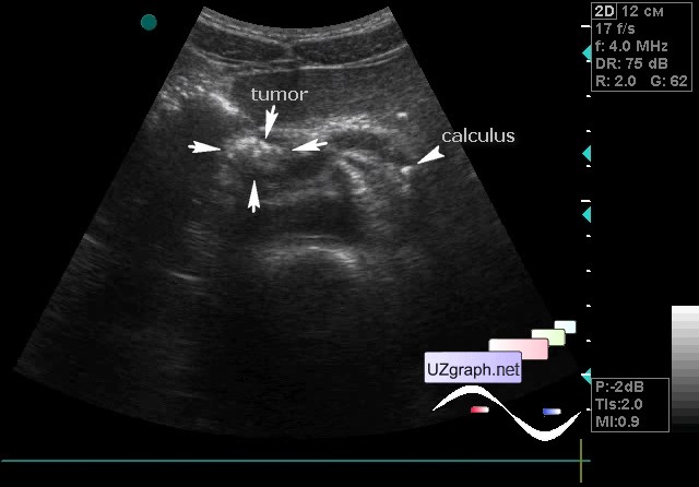
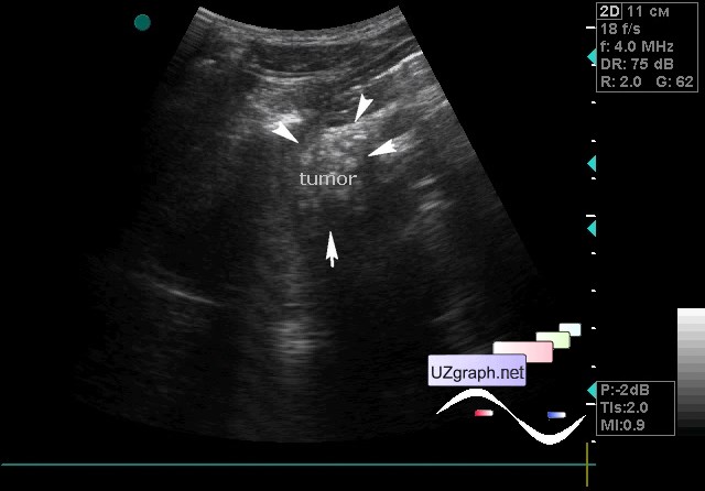
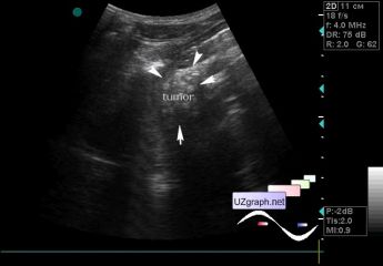
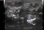
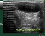
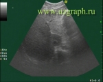
    On the ultrasound the virsung duct is enlarged, in it in the area of the tail of the pancreas there are multiple calculi up to 5 mm. In the head of the pancreas hyperechoic lesion up to 2 cm, with an uneven contour, with an acoustic shadow, a twinkling artifact on the CFM (tumor?).

    Urgent consultation of the surgeon and abdomen CT was recommended.

    external link

:: attachments(4) ::

:: file 1 ::

:: file 2 ::

:: file 3 ::

:: file 4 ::


 Random 
Ultrasound cases - Cranial mass
    

Ultrasound cases - Neonatal hemorrhagic syndrome
    

Ultrasound cases - Condition after testicular surgery - cryptorchidism
    

Ultrasound cases - PDA forth and back
    

Ultrasound cases - Left inferior vena cava
    

 Dictionary 
PASP

HR

PD

BPD

PSV

LVPWd

SV

AC

VTI

EF


 Calculators 
Volume (ellipse formula)

Body surface area

Type of an infant hip joint

Resistive index

Estimated fetal weight by Ultrasound


 Similar topics 
Ultrasound cases - Hirschsprung disease on ultrasound
    

Ultrasound cases - Intestinal Infiltrate
    

Ultrasound cases - Abdominal cryptorchidism
    

Ultrasound cases - Diffuse heterogeneous liver
    

Ultrasound cases - Postoperative abdominal wall fistula
    

Ultrasound cases - Stump appendicitis
    

Ultrasound cases - Liver scar after resection
    

Ultrasound cases - Subdiaphragmatic movable fluid collection (UFO)
    

Ultrasound cases - Lesion of the spleen or the case when it is easy to not notice
    

Ultrasound cases - I’m normal but others ...