Log in    RU  


Main page :: Forum :: Ultrasound cases - Sonograms, cine-loops etc. ::

Thyroid lesion 2


Tags: Thyroid gland sonography, GE Vivid 3, Images, Video, Clinical report, Adult

   Posts
 
  Thyroid lesion 2
#1

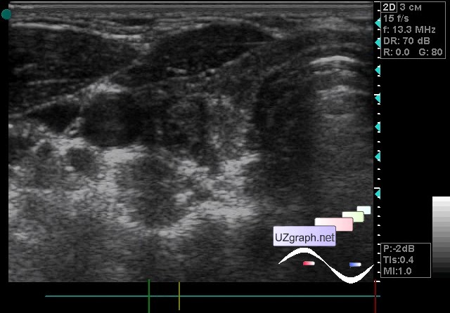
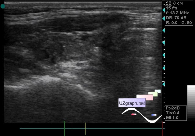
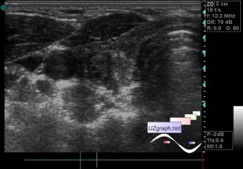
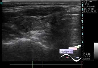
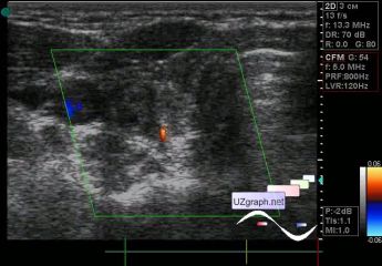
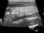
    A 22-year-old patient came for the control ultrasound of the thyroid gland, from her words after a course of radioactive iodine.

    The previous ultrasound report describes the lesion of the right lobe with microcalcinates twice as large in size.

    On the current ultrasound a hypoechoic formation upto 7-8 mm is visualized, at the CFM without blood flow (adenoma? other?).

    Looking closely at the video on a normal monitor (on the device it is a CRT and very faded, can break your eyes) I can see an isoechogenic area with hyperechoic micro inclusions, more medially than this lesion but it's hard for me to say is it a lesion and even with microcalcinates because hyperechoic points are projected strictly on the walls of blood vessels, more like a compressed area of the gland.

    As many doctors - as many opinions.

:: attachments(4) ::

:: file 1 ::

:: file 2 ::

:: file 3 ::

:: file 4 ::


 Random 
Ultrasound cases - Anuria. Acute renal failure
    

Ultrasound cases - Abdominal syndrome & bladder diverticulum
    

Ultrasound cases - Liver scar after resection
    

Ultrasound cases - Subcapsular kidney cysts
    

Ultrasound cases - Condition after choledochal cyst removal
    

 Ad 


 Dictionary 
LVESD

LVESV

PA

AC

CFM

PD

RAP

LVOT

GS

Anatomical M-mode


 Calculators 
Qp:Qs

TI-RADS ACR

Teicholz formula ejection fraction

Thyroid volume

Modified Simpson’s Rule formula (3 disks) ejection fraction


 Similar topics 
Ultrasound cases - Tirads 5 multinodular
    

Ultrasound cases - Pediatric Tirads 5
    

Ultrasound cases - Tirads 5
    

Ultrasound cases - Thyroiditis 2
    

Ultrasound cases - Diffuse-focal thyroid changes, parathyroid glands adenomas
    

Ultrasound cases - Accessory Thyroid Lobe
    

Ultrasound cases - Area with hyperechoic dot inclusions
    

Ultrasound cases - Thyroiditis
    

Ultrasound cases - Hypoplasia of the left thyroid lobe
    

Ultrasound cases - Cystic-solid volume of the thyroid gland