Log in    RU  


Main page :: Forum :: Ultrasound cases - Sonograms, cine-loops etc. ::

Tirads 5 multinodular


Tags: Thyroid gland sonography, GE Logiq E, Clinical report, Pediatric

   Posts
 
  Tirads 5 multinodular
#1

    An 8-year-old child came for a follow-up ultrasound of the thyroid gland regarding a nodule; there was no previous ultrasound report.

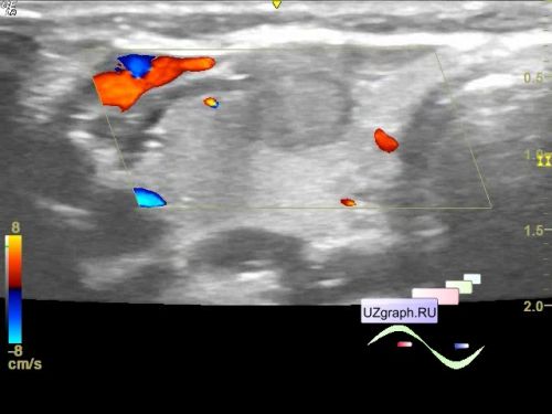
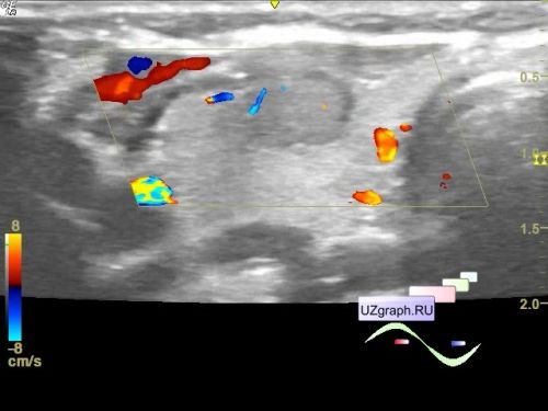
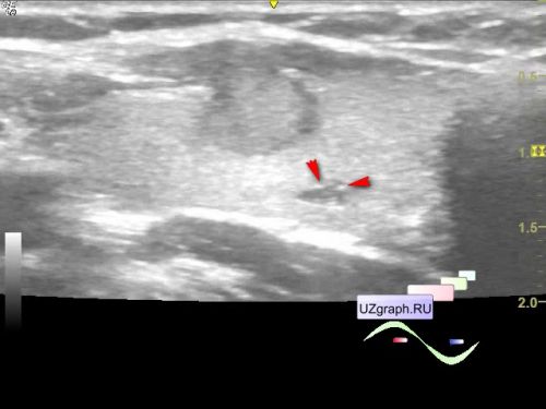
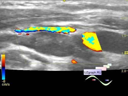
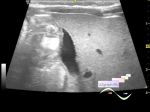
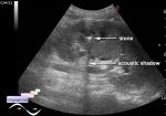
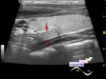
    On ultrasound, 3 lesions are visualized in the right lobe: the largest in the central anterior part of the lobe, isoechoic with a hypoechoic rim, with an uneven, fuzzy contour, especially in the anterior sections, where its border with the muscles is not distinguishable; at CFM with blood flow, along the periphery hyperechoic dot inclusions are visualized (microcalcifications?), size 8x5x9mm (Tirads 5); in the posterior part of the lobe and in the area of the isthmus, hypoechoic lesions with hyperechoic dot inclusions are visualized; at CFM without blood flow, irregular in shape, with an unclear contour, the lesion in the posterior part has a predominantly oblique-vertical orientation, measuring 3x3x6 mm, and the lesion in the isthmus area measuring up to 3x2 mm (Tirads 5).

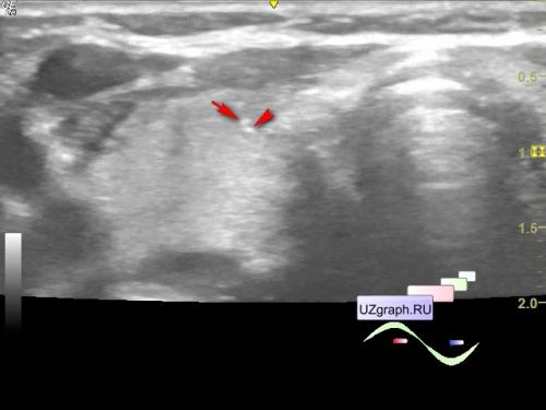
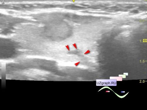
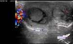
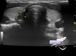
    In the lateral sections of the neck, lymph nodes are visualized, larger on the right, 2 LNs up to 17 mm in size; on the left there are 2 LNs up to 11 mm, at CFM in the LNs on the right there is blood flow in the hilum area.


 Random 
Ultrasound cases - Subcapsular kidney cysts
    

Ultrasound cases - Cavernous Transformation of the Portal Vein
    

Ultrasound cases - Wrist edema
    

Ultrasound cases - Goiter Hashimoto
    

Ultrasound cases - Pneumatosis intestinalis, intussusception etc.
    

 Ad 


 Dictionary 
B-mode

IVSd

PA

DPD

LAP

LVESD

SF

ESD

GS

BSA


 Calculators 
"Bullet" formula (1 disk) ejection fraction

Modified Simpson’s Rule formula (3 disks) ejection fraction

Modified Simpson’s Rule formula (2 disks) ejection fraction

Type of an infant hip joint

Area-length (Simpson) formula ejection fraction


 Similar topics 
Ultrasound cases - Pediatric Tirads 5
    

Ultrasound cases - Tirads 5
    

Ultrasound cases - Thyroiditis 2
    

Ultrasound cases - Diffuse-focal thyroid changes, parathyroid glands adenomas
    

Ultrasound cases - Accessory Thyroid Lobe
    

Ultrasound cases - Area with hyperechoic dot inclusions
    

Ultrasound cases - Thyroiditis
    

Ultrasound cases - Hypoplasia of the left thyroid lobe
    

Ultrasound cases - Cystic-solid volume of the thyroid gland
    

Ultrasound cases - Hyperparathyroidism?