Log in    RU  


Main page :: Forum :: Ultrasound cases - Sonograms, cine-loops etc. ::

Pediatric Tirads 5


Tags: Thyroid gland sonography, GE Logiq E, Clinical report, Pediatric

   Posts
 
  Pediatric Tirads 5
#1

    A 4-year-old child was referred for ultrasound by an endocrinologist.

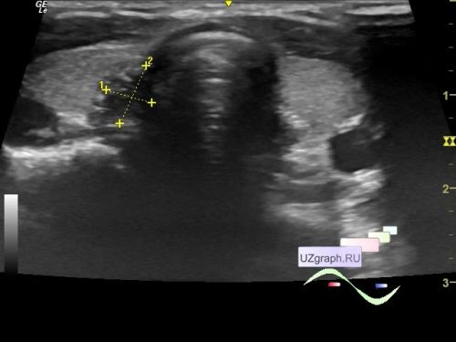
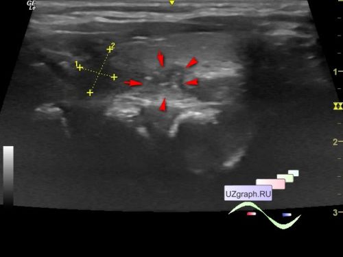
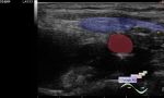
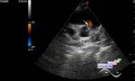
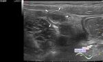
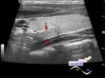
    On ultrasound in the medial part of the right lobe, a hypoechoic lesion with hyperechoic dot inclusions with a fuzzy uneven contour, 8x6x5 mm in size (Tirads 5, differential diagnosis: thymus ectopia, etc.) is visualized intimately located to the trachea and esophagus, on the CFM the blood flow along the contour.

    PS. The calipers on the photo and video are a GE scanner glitch.


 Random 
Ultrasound cases - Unicornuate uterus with rudimentary horn
    

Ultrasound cases - Subhepatic appendicitis and omentitis
    

Ultrasound cases - Starry sky sign
    

Ultrasound cases - Right hypogastrium mass
    

Admin room - Who are Sonologists?

 Dictionary 
TAV

FL

PG

EFW

Anatomical M-mode

Ved

ESD

SF

LVEDD

FNA


 Calculators 
Pulmonary artery pressure

Volume (ellipse formula)

Teicholz systolic cardiac function

Pressure gradient

Gestational age by Ultrasound


 Similar topics 
Ultrasound cases - Tirads 5 multinodular
    

Ultrasound cases - Tirads 5
    

Ultrasound cases - Thyroiditis 2
    

Ultrasound cases - Diffuse-focal thyroid changes, parathyroid glands adenomas
    

Ultrasound cases - Accessory Thyroid Lobe
    

Ultrasound cases - Area with hyperechoic dot inclusions
    

Ultrasound cases - Thyroiditis
    

Ultrasound cases - Hypoplasia of the left thyroid lobe
    

Ultrasound cases - Cystic-solid volume of the thyroid gland
    

Ultrasound cases - Hyperparathyroidism?