Log in    RU  


Main page :: Forum :: Ultrasound cases - Sonograms, cine-loops etc. ::

Tirads 5


Tags: Thyroid gland sonography, GE Logiq E, Clinical report, Pediatric

   Posts
 
  Tirads 5
#1

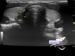
    A 7-year-old child was sent for a thyroid ultrasound due to obesity.

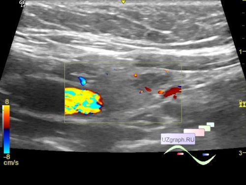
    In the right lobe, a hypoechoic lesion with an uneven, clear contour and echogenic dot microinclusions is visualized; on the color flow map, the blood flow along the contour, 5 x 11 x 3 mm in size, oblique-horizontal orientation (TiRads 5 (9 points) ).

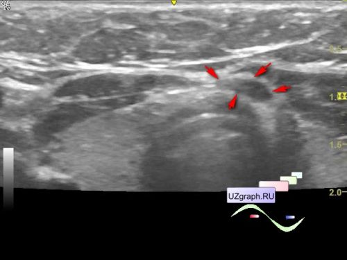
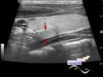
    An an/hypoechoic rounded inclusion up to 5 x 2 x 4 mm is visualized in the isthmus region on the left, without blood flow at CFM (differential diagnosis: pretracheal lymph node, thyroid node, etc.)


 Random 
Ultrasound cases - Thymic lesion
    

Ultrasound cases - Calcific abdominal lymphadenopathy
    

Scientific - Playlist Bones ultrasound

Ultrasound cases - Where is the testicle?
    

Ultrasound cases - Chest lesion
    

 Dictionary 
Ved

IVC

Anatomical M-mode

LN

PA

DPD

Color M-mode

PW

ESV

TAV


 Calculators 
Estimated fetal weight by Ultrasound

Myocardial mass

TI-RADS

Pulmonary artery pressure

Body surface area


 Similar topics 
Ultrasound cases - Tirads 5 multinodular
    

Ultrasound cases - Pediatric Tirads 5
    

Ultrasound cases - Thyroiditis 2
    

Ultrasound cases - Diffuse-focal thyroid changes, parathyroid glands adenomas
    

Ultrasound cases - Accessory Thyroid Lobe
    

Ultrasound cases - Area with hyperechoic dot inclusions
    

Ultrasound cases - Thyroiditis
    

Ultrasound cases - Hypoplasia of the left thyroid lobe
    

Ultrasound cases - Cystic-solid volume of the thyroid gland
    

Ultrasound cases - Hyperparathyroidism?