Log in    RU  


Main page :: Forum :: Ultrasound cases - Sonograms, cine-loops etc. ::

Accessory Thyroid Lobe


Tags: Thyroid gland sonography, GE Logiq E, Clinical report, Pediatric

   Posts
 
  Accessory Thyroid Lobe
#1

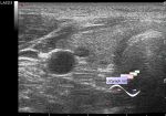
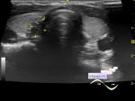
    A teenager came to the public clinic for an ultrasound of the thyroid gland.

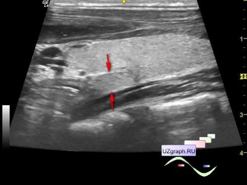
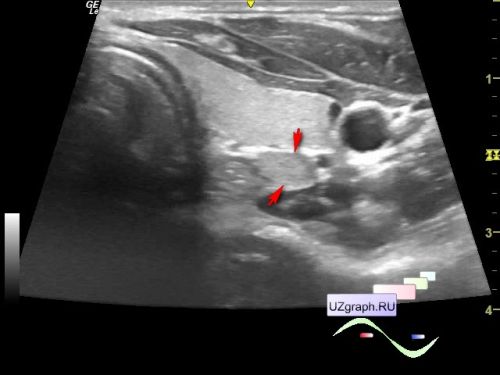
    On ultrasound, an accessory lobe is visualized in the posterior parts of the left lobe.

    I found 2 similar echograms in 3 (2 of them are reissues) russian sonography manuals.

    Few quotes in my translation:

    "Anatomical variant of the thyroid gland structure in a healthy 12-year-old child. Oblique-longitudinal scanning of the left lobe. An accessory lobule of the gland is determined along the posterior surface of the lobe, in the region of its lower pole."

    Clinical Guide to Ultrasound Diagnostics in Pediatrics, under ed. of M.I. Pykov, 1998, p. 332, fig. 6

    Children's ultrasound diagnostics. under ed. of M.I. Pykov, 2001, p. 570, fig. 6

    "longitudinal view of the accessory lateral lobule of the thyroid gland"

    A practical guide to ultrasound diagnostics. General ultrasound diagnostics. under ed. of V.V. Mitkov, 2006, p. 613, fig. 16

    And one more quote for a general excursion into the topic:

    "Other anomalies in the development of the thyroid gland are the accessory lobes. The most common is the third, pyramidal lobe. At echography, its determined anteriorly and cranial to the isthmus. The accessory lobes may have a different location."

    Clinical guide to ultrasound diagnostics. under ed. of V.V. Mitkov, Volume 2, 1996, p. 379


 Random 
Ultrasound cases - Colloid nodules
    

Ultrasound cases - Condition after surgery for varicocele
    

On-call room - Web-Tools for sonography: Online Calculators!
    

Scientific - Inguinal hernia in a child sonography
    

Ultrasound cases - Venous lacunae
    

 Ad 


 Dictionary 
TAV

AC

SF

GA

FVI

Qp-Qs

LVAs

LN

MC

HR


 Calculators 
Volume (ellipse formula)

Modified Simpson’s Rule formula (2 disks) ejection fraction

Resistive index

Myocardial mass

Pressure gradient


 Similar topics 
Ultrasound cases - Tirads 5 multinodular
    

Ultrasound cases - Pediatric Tirads 5
    

Ultrasound cases - Tirads 5
    

Ultrasound cases - Thyroiditis 2
    

Ultrasound cases - Diffuse-focal thyroid changes, parathyroid glands adenomas
    

Ultrasound cases - Area with hyperechoic dot inclusions
    

Ultrasound cases - Thyroiditis
    

Ultrasound cases - Hypoplasia of the left thyroid lobe
    

Ultrasound cases - Cystic-solid volume of the thyroid gland
    

Ultrasound cases - Hyperparathyroidism?