Log in    RU  


Main page :: Forum :: Ultrasound cases - Sonograms, cine-loops etc. ::

Cyst of the epididymis and appendages


Tags: Scrotum sonography, GE Vivid 3, Images, Video, Clinical report, Adult

   Posts
 
  Cyst of the epididymis and appendag...
#1

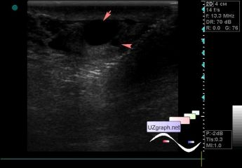
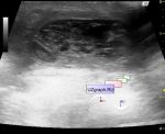
    A 21-year-old patient came to the scrotum ultrasound with complaints of intermittent pain in the scrotum and palpable lesion in the right half of the scrotum.

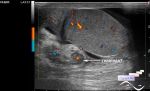
    On an ultrasound in the right half of the scrotum upto 1 ml of free fluid, side to the testicle in the projection of the epididymis an anechoic oval shape lesion type of a cyst (epididymis cyst?) up to 1 cm is visualized, and another one is up to 2 mm (testicular appendix).

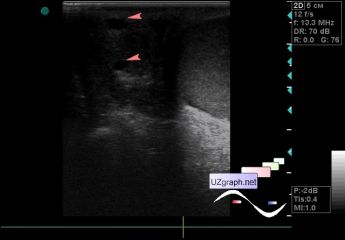
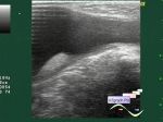
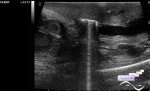
    In the other half of the scrotum there are 3 more similar inclusions in the projection of the epididymis head (testicular appendages?)

:: attachments(3) ::

:: file 1 ::

:: file 2 ::

:: file 3 ::


 Random 
Ultrasound cases - Parathyroid lesions
    

Admin room - Flash end of life

Ultrasound cases - Epididymis cysts
    

On-call room - Domo arigato mr. Roboto

Ultrasound cases - Cholelithiasis plus 3
    

 Dictionary 
LVESD

CRL

Ved

MSD

FNA

LAP

FL

CW

EDV

AC


 Calculators 
Pulmonary artery pressure

Qp:Qs

Type of an infant hip joint

Resistive index

Pressure gradient


 Similar topics 
Ultrasound cases - Abdominal cryptorchidism
    

Ultrasound cases - Neonatal testicular torsion
    

Ultrasound cases - Fibrinous epididymitis
    

Ultrasound cases - Scrotum hematocele
    

Ultrasound cases - Varicocele in a child
    

Ultrasound cases - Hydrocele (dropsy of the scrotum) in a child
    

Ultrasound cases - Air in the scrotum, post surgery condition
    

Ultrasound cases - Multi-chamber scrotum cyst
    

Ultrasound cases - Diffuse changes of the testicle in a teenager (orchitis)
    

Ultrasound cases - Thickening of the spermatic cord in a teenager