Log in    RU  


Main page :: Forum :: Ultrasound cases - Sonograms, cine-loops etc. ::

Regional cervical lymphadenitis


Tags: Soft tissues sonography, GE Vivid 3, Images, Video, Clinical report, Adult

   Posts
 
  Regional cervical lymphadenitis
#1

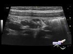
    A 19-year-old patient came to an ultrasound scan of the LN, in the region of the middle third of the lateral surface of the neck a dense lesion is palpated, and soft tissue is thickened in the region of the mandibular angle on the same side.

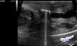
    On ultrasound in the indicated areas, enlarged LNs are visualized, without blood flow at the CFM (regional cervical lymphadenitis).

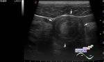
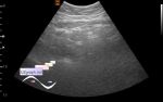
    Separate interest was aroused by the semilunar regions below the rounded LNs in the area of mandibular angle, which at first aroused suspicion for abscessing, but still I came to the conclusion that this is a LNs but just with an unusual shape.

:: attachments(2) ::

:: file 1 ::

:: file 2 ::

  12:08 10-10-2018
 
#2

    During the next visit the patient said that before the occurrence of this situation, she was resting on the sea, then the temperature and these bumps at her neck appeared.


 Random 
Ultrasound cases - Neonatal mastitis
    

Ultrasound cases - Partial tears of the lateral meniscus
    

Ultrasound cases - Periventricular hyperechogenicity
    

Ultrasound cases - Wrist abscess
    

Ultrasound cases - Hyperparathyroidism?


 Dictionary 
Aliasing doppler artifact

SF

LVEDV

RAP

Anatomical M-mode

CRL

CW

Gain

HC

Ved


 Calculators 
Thyroid volume

Pressure gradient

Volume (ellipse formula)

Body surface area

TI-RADS


 Similar topics 
Ultrasound cases - Penetrating injury of the femur
    

Ultrasound cases - Cephalohematoma
    

Ultrasound cases - Urachus fistula
    

Ultrasound cases - Cranial mass
    

Ultrasound cases - Pilonidal coccyx cyst
    

Ultrasound cases - Palm fistula, in search of pencil lead
    

Ultrasound cases - Postoperative abdominal wall fistula
    

Ultrasound cases - Nucks cyst
    

Ultrasound cases - Paraproctitis
    

Ultrasound cases - Submandibular area lesion