Log in    RU  


Main page :: Forum :: Ultrasound cases - Sonograms, cine-loops etc. ::

Infected Medial Umbilical Ligament


Tags: Abdomen sonography, Soft tissues sonography, GE Versana, Clinical report, Pediatric

   Posts
 
  Infected Medial Umbilical Ligament
#1

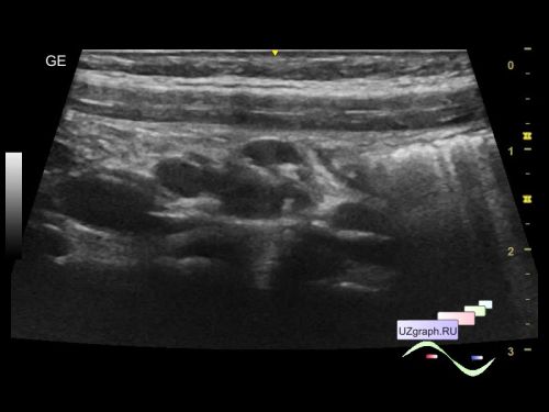
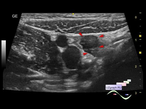
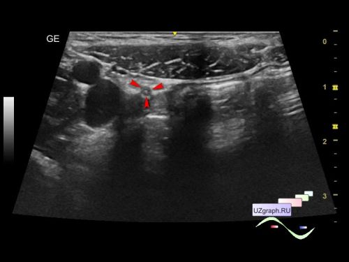
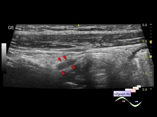
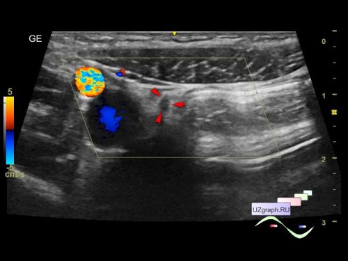
    A 6-year-old child recently underwent a laparoscopic appendectomy, came for a follow-up abdominal ultrasound due to sludge in the gallbladder that was detected earlier.

    On ultrasound, the gallbladder is without features, pronounced mesadenitis is visualized, and in the projection of the postoperative area, an additional tubular hypoechoic structure with a hyperechoic center is visualized extending from the anterior abdominal wall near the navel to the small pelvis, without blood flow on the CDI, up to 2 mm in diameter, a small amount of free fluid (differential diagnosis: fistula / infected medial umbilical ligament, etc.).

    An urgent consultation with a surgeon is recommended.

    Quotes:

    "The medial umbilical folds are bilateral raised ridges of parietal peritoneum in the deep aspect of the anterior abdominal wall formed by the underlying medial umbilical ligaments running from the pelvis to the umbilicus. The medial umbilical ligaments are anatomical remnants of the obliterated foetal umbilical arteries.

    ...

    The medial umbilical folds are variably seen on CT depending on the density of the ligament and the amount of perivesical and intraperitoneal fat. These folds can be displaced laterally by a large amount of ascites."

    external link

    "Related to the urinary bladder, anteriorly there are the following folds:

    one median umbilical fold on the median umbilical ligament (which in turn, contains the urachus)

    two medial umbilical folds on the occluded umbilical artery

    two lateral umbilical folds on the inferior epigastric vessels"

    external link

    external link

    "After the descent of bladder into the pelvis, the apical part narrows to form epithelialized fibromuscular remnant, the median umbilical ligament or the urachus. It lies in the extraperitoneal space between the fascia transversalis and parietal peritoneum along with the medial umbilical ligaments (formed from obliterated umbilical arteries). Usually, it is located in the midline. Occasionally, it may merge with the medial ligaments and may be deviated towards right or left of midline."

    external link

    "The removal of the urachal remnant and medial umbilical ligaments was completed with a median operative time of 92 min (range 69–128).

    ...

    As medial umbilical ligaments can develop chronic infection in the event of omphalitis, the medial umbilical ligaments were resected continuously."

    external link

    "Treatment of urachus anomalies requires removing the urachus throughout its entire length including each medial umbilical ligament as well as the associated peritoneum."

    external link

    "Patients were treated according to the following protocol: transumbilical access with one 10 mm port using the laparoscope with working channel. The appendix was mobilized and delivered through the umbilical port and tied extracorporeally and removed."

    external link

    In other words, with laparoscopic appendectomy, infection of the medial umbilical ligaments is possible ...


 Random 
Ultrasound cases - Polyposis of the gallbladder
    

On-call room - MRI communism

Ultrasound cases - Mirror artifact vs collapsed lung
    

Ultrasound cases - Accessory thymic lobe
    

Ultrasound cases - Increased blood flow in the thyroid
    

 Dictionary 
PW

Freeze

CW

LN

SV

CFM

AC

PADP

B-mode

ESD


 Calculators 
Pressure gradient

Estimated fetal weight by Ultrasound

Thyroid volume

Pulmonary artery pressure

Teicholz systolic cardiac function


 Similar topics 
Ultrasound cases - Portal hypertension
    

Ultrasound cases - Lesion in the projection of the adrenal gland 2
    

Ultrasound cases - Gallbladder, saw sign
    

Ultrasound cases - Ileotiphlitis and mesadenitis
    

Ultrasound cases - Cholelithiasis and hyperechoic gallbladder wall
    

Ultrasound cases - Portal vein spontaneous enhancement (mononucleosis)
    

Ultrasound cases - Hypoechoic spleen lesion
    

Ultrasound cases - Acute Appendicitis, periappendicular infiltrate
    

Ultrasound cases - Cavernous Transformation of the Portal Vein
    

Ultrasound cases - Diffuse-focal changes in the liver, thick left kidney