Log in    RU  


Main page :: Forum :: Ultrasound cases - Sonograms, cine-loops etc. ::

Birads 4


Tags: Breast sonography, GE Vivid 3, Images, Video, Clinical report, Adult

   Posts
 
  Birads 4
#1

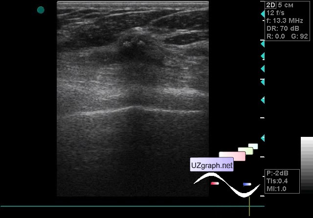
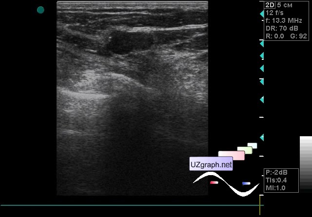
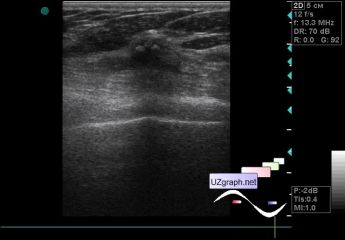
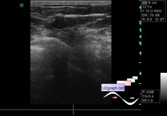
    A 57-year-old patient came to a screening ultrasound of the mammary glands; earlier no lesions were detected in the mammary gland.

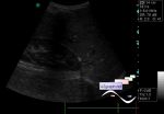
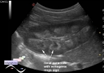
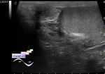
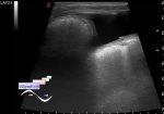
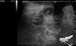
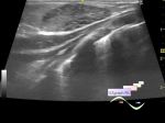
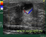
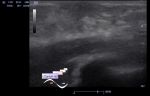
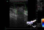
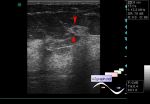
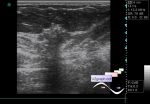
    At ultrasound scan in the outer quadrants of the left breast shows an irregular oval shaped hypoechoic lesion, with hyperechoic microinclusions (microcalcinates?), horizontal orientation, on the CFM without blood flow, upto 1cm in size (birads 4?).

:: attachments(4) ::

:: file 1 ::

:: file 2 ::

:: file 3 ::

:: file 4 ::


 Random 
Ultrasound cases - Bilateral popliteal cysts with legs
    

Ultrasound cases - Purulent / Suppurative lymphadenitis
    

Ultrasound cases - Pneumonia
    

Ultrasound cases - Ovarian apoplexy 3
    

Ultrasound cases - Isolated spleen calcification
    

 Dictionary 
RVOT

BPD

TI-RADS

LVESD

TVUS

IVSd

CW

PRF

EDD

EF


 Calculators 
Type of an infant hip joint

TI-RADS

Teicholz systolic cardiac function

Estimated fetal weight by Ultrasound

Gestational age by Ultrasound


 Similar topics 
Ultrasound cases - Mastitis and cysts
    

Ultrasound cases - Neonatal mastitis
    

Ultrasound cases - Adolescent Gynecomastia
    

Ultrasound cases - Subacute mastitis (cyst with inflammation) in a teenager
    

Ultrasound cases - Breast Cysts
    

Ultrasound cases - Infant isolated thelarche
    

Ultrasound cases - Another Birads 4-5
    

Ultrasound cases - Star-like lesion
    

Ultrasound cases - Birads 4-5
    

Ultrasound cases - Breast lesions 3