Log in    RU  


Main page :: Forum :: Ultrasound cases - Sonograms, cine-loops etc. ::

Return of spermatocele


Tags: Scrotum sonography, Images, Video, Clinical report, Esaote MyLab 70, Pediatric

   Posts
 
  Return of spermatocele
#1

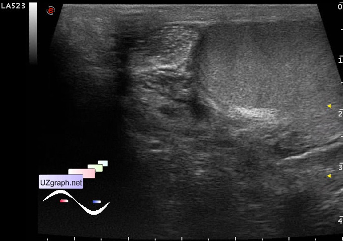
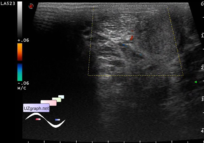
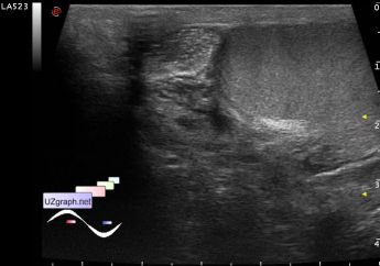
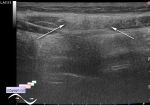
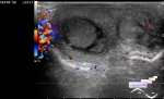
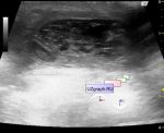
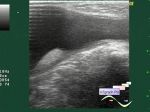
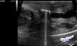
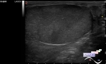
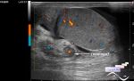
    Teenager 14 years-old has surgery of spermatocele removal in history, at control ultrasonography in the projection of the head of the epididymis visualized oval-shaped mass with hyperechoic microlesions inside up to 1,5x1 cm without blood flow at CFM.

    external link

:: attachments(4) ::

:: file 1 ::

:: file 2 ::

:: file 3 ::

:: file 4 ::


 Random 
Ultrasound cases - Septum pellucidum agenesis
    

Scientific - Acute abdomen in a child sonography
    

Ultrasound cases - Traumatic epididymitis
    

Ultrasound cases - Lesions in the projection of the adrenal glands
    

Ultrasound cases - Subdural hemorrhage 2
    

 Dictionary 
PA

TAV

PASP

GS

AC

RAP

GA

PADP

PG

FL


 Calculators 
Pulmonary artery pressure

Gestational age by Ultrasound

Resistive index

Thyroid volume

Volume (ellipse formula)


 Similar topics 
Ultrasound cases - Abdominal cryptorchidism
    

Ultrasound cases - Neonatal testicular torsion
    

Ultrasound cases - Fibrinous epididymitis
    

Ultrasound cases - Scrotum hematocele
    

Ultrasound cases - Varicocele in a child
    

Ultrasound cases - Hydrocele (dropsy of the scrotum) in a child
    

Ultrasound cases - Air in the scrotum, post surgery condition
    

Ultrasound cases - Multi-chamber scrotum cyst
    

Ultrasound cases - Diffuse changes of the testicle in a teenager (orchitis)
    

Ultrasound cases - Thickening of the spermatic cord in a teenager