Log in    RU  


Main page :: Forum :: Ultrasound cases - Sonograms, cine-loops etc. ::

Split-image artifact


Tags: Abdomen sonography, GE Vivid 3, Images, Video, Clinical report, Adult

   Posts
 
  Split-image artifact
#1

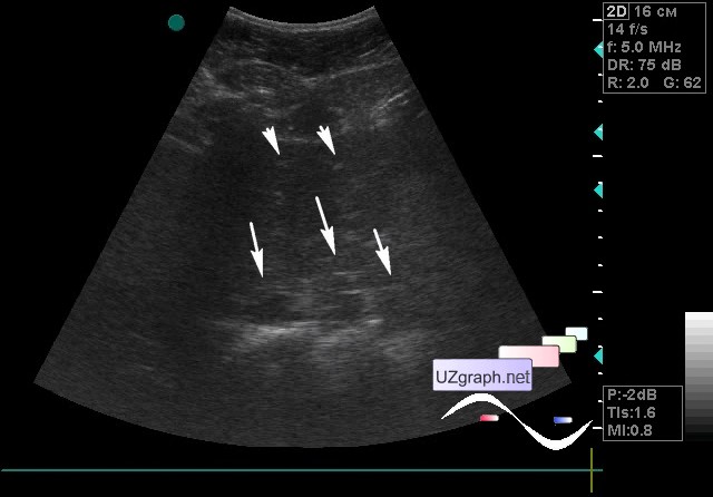
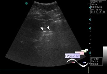
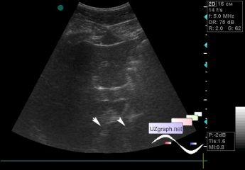
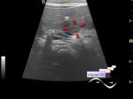
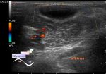
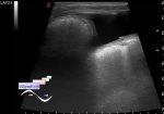
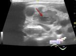
    A 22-year-old patient came for an abdomen ultrasound.

    Object reproduction artifact or refractive artifact (ie, caused by partial refraction - partial distortion of the direction / refraction of the beam after it passes the boundary of environments with different densities, as a result, the main and additional false images are obtained), a similar artifact was described with doubling of the uterus, located behind the filled bladder (split-image artifact) - external link

    And here the anterior abdominal wall is probably the refracting boundary of the environments, since we see a doubling of the liver, vertebra, gas in the intestines, and even the tripling of the pancreatic head.

    external link

:: attachments(4) ::

:: file 1 ::

:: file 2 ::

:: file 3 ::

:: file 4 ::


 Random 
Ultrasound cases - Palm fistula, in search of pencil lead
    

Ultrasound cases - Myxomatous degeneration of MV
    

Ultrasound cases - Condition after diaphragmatic hernia
    

Ultrasound cases - Single doubled kidney and Mullerian duct cyst in the infant
    

Ultrasound cases - Subcutaneous lesion above the sternum
    

 Dictionary 
ESV

CW

Gain

PA

DPD

TV

SV

GS

LMP

PASP


 Calculators 
Volume (ellipse formula)

Type of an infant hip joint

TI-RADS

Resistive index

Pressure gradient


 Similar topics 
Ultrasound cases - Hirschsprung disease on ultrasound
    

Ultrasound cases - Intestinal Infiltrate
    

Ultrasound cases - Abdominal cryptorchidism
    

Ultrasound cases - Diffuse heterogeneous liver
    

Ultrasound cases - Postoperative abdominal wall fistula
    

Ultrasound cases - Stump appendicitis
    

Ultrasound cases - Liver scar after resection
    

Ultrasound cases - Subdiaphragmatic movable fluid collection (UFO)
    

Ultrasound cases - Lesion of the spleen or the case when it is easy to not notice
    

Ultrasound cases - I’m normal but others ...