Log in    RU  


Main page :: Forum :: Ultrasound cases - Sonograms, cine-loops etc. ::

Lesion in the projection of the pancreas body


Tags: Abdomen sonography, GE Logiq E, Clinical report, Pediatric

   Posts
 
  Lesion in the projection of the pan...
#1

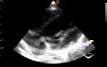
    A 1-year-old child was sent to the public clinic for an abdominal ultrasound, according to the accompanying person, he had previously had some kind of infectious disease.

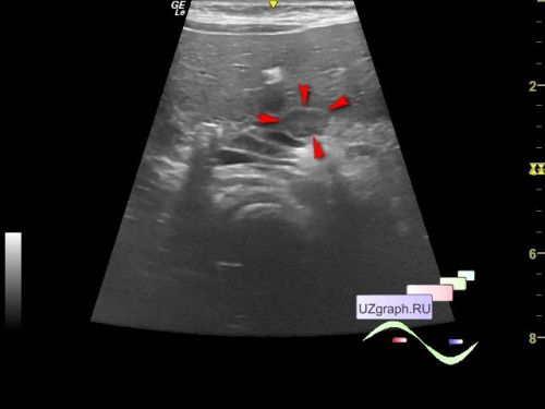
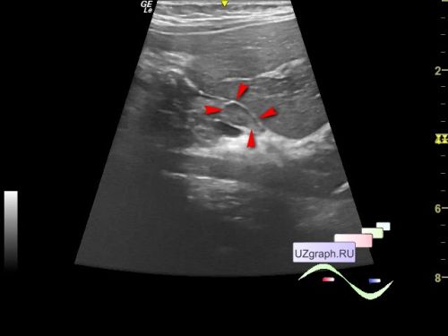
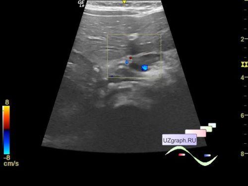
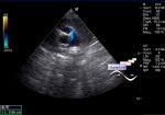
    In the projection of the pancreas body, a hypoechoic oval inclusion is visualized, without blood flow on CFM, up to 11 x 6 mm in size (differential diagnosis: lymph node, etc.)

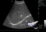
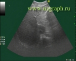
    In the mesogastrium, more than 2 lymph nodes up to 6 mm are also visualized (differential diagnosis: mesadenitis, etc.)


    external link


 Random 
Ultrasound cases - Neuroblastoma. Diffuse infiltration of liver
    

Ultrasound cases - Doubling of the kidney
    

Ultrasound cases - Abdominal syndrome, two in one
    

Ultrasound cases - Good vs Evil
    

Ultrasound cases - Snowball
    

 Dictionary 
PG

Qp-Qs

Color M-mode

GA

TAMEAN

CFM

TAV

FL

LVESV

ESV


 Calculators 
Volume (ellipse formula)

Resistive index

Thyroid volume

Pressure gradient

TI-RADS


 Similar topics 
Ultrasound cases - Hirschsprung disease on ultrasound
    

Ultrasound cases - Intestinal Infiltrate
    

Ultrasound cases - Abdominal cryptorchidism
    

Ultrasound cases - Diffuse heterogeneous liver
    

Ultrasound cases - Postoperative abdominal wall fistula
    

Ultrasound cases - Stump appendicitis
    

Ultrasound cases - Liver scar after resection
    

Ultrasound cases - Subdiaphragmatic movable fluid collection (UFO)
    

Ultrasound cases - Lesion of the spleen or the case when it is easy to not notice
    

Ultrasound cases - I’m normal but others ...