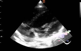
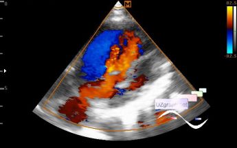
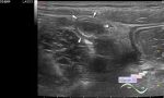
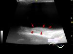
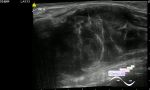
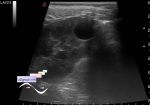
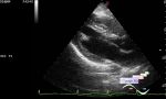
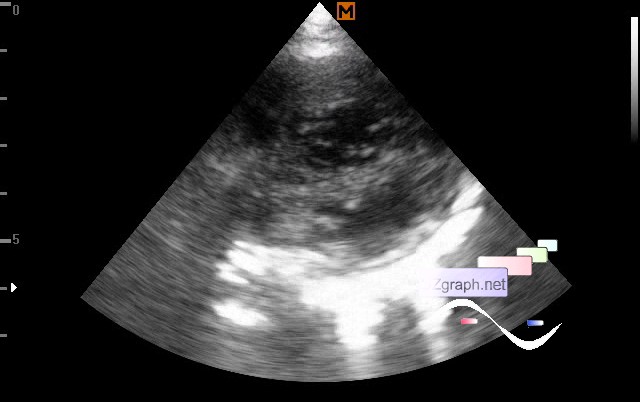
Left ventricular non-compaction
Tags: Cardiac sonography(Echocardiography), Images, Video, Clinical report, Medison Sonoace R7, Pediatric
| Posts | |||
| Left ventricular non-compaction | #1 |
| |||||
:: file 1 ::
 :: file 2 ::
 :: file 3 ::
 :: file 4 ::
 :: file 5 ::
 :: file 6 :: | |||||