Child 9 years-old in the ICU of the Children's City Clinical Hospital, with cachexia, bedsore ... the main diagnosis is chylopericardium, pericardiectomy.
A follow-up ultrasound scan was prescribed for fluid in the pleural cavities. According to the treating person the other day, chest CT scan was performed, on which they could not exclude (expressed suspicion) the presence of dense liquid (like "porridge") contents of one of the halves of the chest, which most likely could not be aspirated.
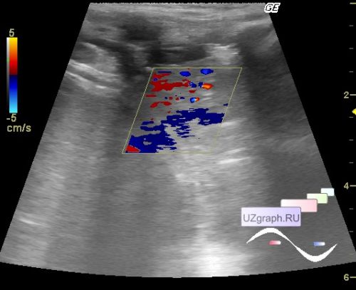
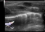
On ultrasound, above the corresponding hemidiaphragm, there is a thin anechoic strip (fluid), above which there is a solid structure, to exclude artifacts, I climbed a rib higher and got the same picture, at CFM with blood flow - an airless lung (hepatization, differential diagnosis: atelectasis, pneumonia, etc.).
1 month after, the child was fattened, but the clinical condition is consistently severe, a follow-up ultrasound scan was prescribed for emergency indications ...
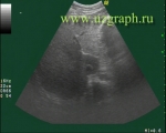
On ultrasound, about 200 ml of free fluid in the small pelvis, visible bowel loops without reliable signs of peristalsis, the rest of the abdominal cavity with a reverberation effect.
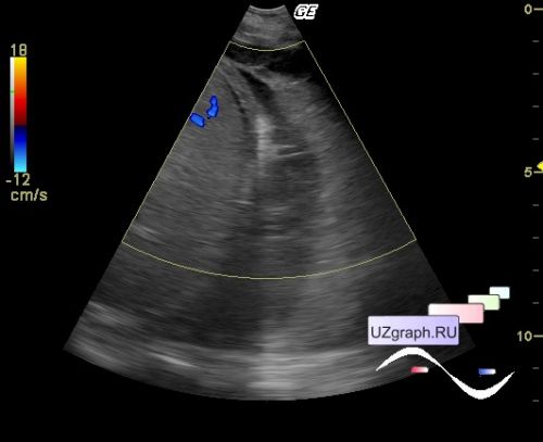
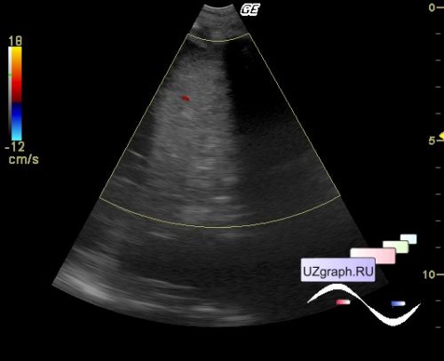
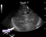
In the pleural: cavity on the right, an airless lung remains, on the left, the lung, according to an ICU doctor, hypoplastic, most of the left half of the chest is occupied by the heart (cardiomegaly according to X-ray), in the projection of the apex of the heart, free fluid (chylopericardium), in the lateral parts of the left chest B, C lines are visualized without reliable mobility on the background of a pronounced excursion (respiratory movements ) chest (differential diagnosis: edema, hypoventilation, pneumonia, pneumothorax, etc.)