Log in    RU  


Main page :: Forum :: Ultrasound cases - Sonograms, cine-loops etc. ::

Adolescent Gynecomastia


Tags: Breast sonography, GE Logiq E, Clinical report, Pediatric

   Posts
 
  Adolescent Gynecomastia
#1

    A 16-year-old teenager was referred by an endocrinologist to a public clinic for an ultrasound scan, including of the mammary glands, with complaints of enlargement of one of the glands.

    According to the teenager, he had previously had a similar situation with another gland, which then returned to normal.

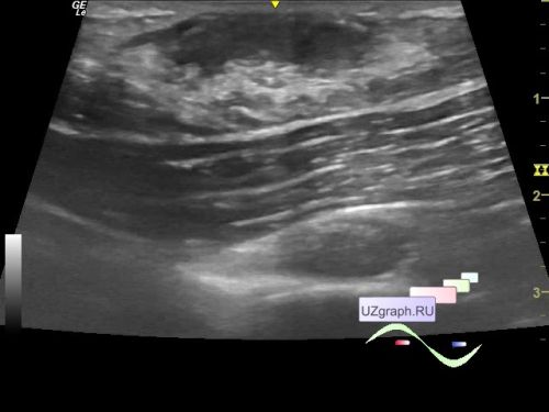
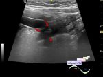
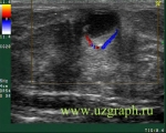
    On ultrasound in the projection of the enlarged mammary gland, a pronounced layer of glandular tissue (gynecomastia) is visualized, without increased blood flow at CFM.

    "Gynecomastia refers to an enlargement of the male breast caused by benign proliferation of the glands ducts and stromal components including fat. It is the most common form of breast swelling seen in adolescent males. During pubertal development, gynecomastia can develop as a result of transient relative imbalances between androgens and estrogens. Pubertal gynecomastia is self-limited in 75 to 90% of adolescents and regresses over 1 to 3 years."

    external link


    external link


 Random 
Ultrasound cases - Thread-like structure in RA
    

Ultrasound cases - nabothian cyst
    

Ultrasound cases - Hirschsprung disease on ultrasound
    

Ultrasound cases - Anechogenic inclusion in the posterior cranial fossa
    

Ultrasound cases - Hirschsprung's
disease after surgery


 Dictionary 
EDV

LMP

Aliasing doppler artifact

LVEDD

LVESV

LVPWd

TI-RADS

EDD

RAP

AC


 Calculators 
Type of an infant hip joint

Pulmonary artery pressure

Body surface area

Thyroid volume

Qp:Qs


 Similar topics 
Ultrasound cases - Mastitis and cysts
    

Ultrasound cases - Neonatal mastitis
    

Ultrasound cases - Subacute mastitis (cyst with inflammation) in a teenager
    

Ultrasound cases - Breast Cysts
    

Ultrasound cases - Infant isolated thelarche
    

Ultrasound cases - Another Birads 4-5
    

Ultrasound cases - Star-like lesion
    

Ultrasound cases - Birads 4-5
    

Ultrasound cases - Breast lesions 3
    

Ultrasound cases - Milky ducts