Log in    RU  


Main page :: Forum :: Ultrasound cases - Sonograms, cine-loops etc. ::

Breast lesions 3


Tags: Breast sonography, GE Vivid 3, Images, Video, Clinical report, Adult

   Posts
 
  Breast lesions 3
#1

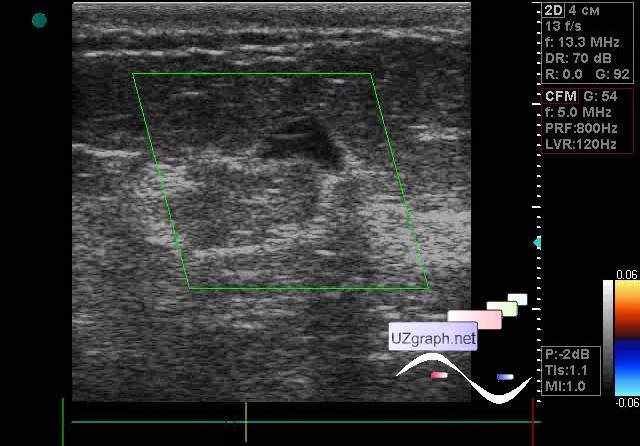
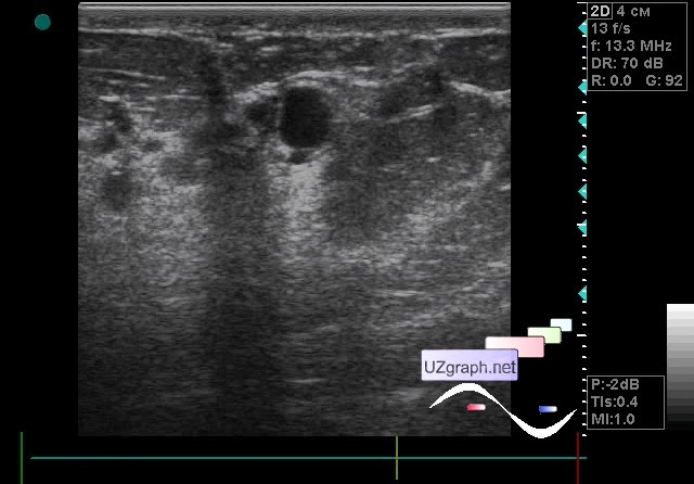
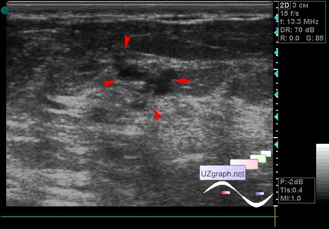
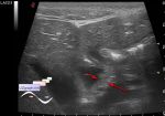
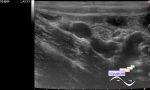
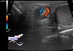
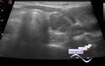
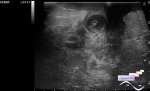
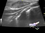
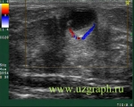
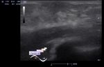
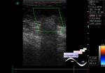
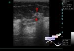
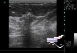
    The patient 45 years old, came to breast screening.

    In the right mammary gland at 5 o' clock anechoic lesion of an irregular oval shape such as a boomerang is visualized, on the CFM without blood flow, up to 9 x 3 x 10 mm in size (cyst?).

    In the left mammary gland several lesions are visualized:

    1) at 11 o' clock anechoic round lesion, without blood flow at CFM, up to 8 x 14 x 11 mm (cyst?);

    2) another 2-3 similar to the previous lesion: the largest at 8 o' clock to 6 x 5 x 5 mm and at 3 o' clock 1-2 another - up to 4 x 3mm each(cysts?);

    3) Also, an an- / hypoechoic lesion of an irregular shape with a fuzzy uneven contour with a tendency to vertical orientation is visualized at 3 o' clock, on the CFM without blood flow, approximately upto (it is not possible to view the longitudinal axis of lesion in one image, to measure it) 9 x 6 x 16 mm (birads 4-5).

    external link

:: attachments(5) ::

:: file 1 ::

:: file 2 ::

:: file 3 ::

:: file 4 ::

:: file 5 ::


 Random 
Ultrasound cases - Strangulated inguinal hernia with necrosis/hematoma
    

Ultrasound cases - Sports interest
    

Ultrasound cases - Giant mass in the female pelvis
    

Ultrasound cases - Choledochal cyst
    

Ultrasound cases - Lesion posterior to the thyroid right lobe
    

 Dictionary 
PG

DPD

ESD

AC

M-mode

BPD

GA

BSA

SV

Gain


 Calculators 
Pressure gradient

Gestational age by Ultrasound

TI-RADS

Resistive index

Volume (ellipse formula)


 Similar topics 
Ultrasound cases - Mastitis and cysts
    

Ultrasound cases - Neonatal mastitis
    

Ultrasound cases - Adolescent Gynecomastia
    

Ultrasound cases - Subacute mastitis (cyst with inflammation) in a teenager
    

Ultrasound cases - Breast Cysts
    

Ultrasound cases - Infant isolated thelarche
    

Ultrasound cases - Another Birads 4-5
    

Ultrasound cases - Star-like lesion
    

Ultrasound cases - Birads 4-5
    

Ultrasound cases - Milky ducts