Log in    RU  


Main page :: Forum :: Ultrasound cases - Sonograms, cine-loops etc. ::

RLQ phlebectasia


Tags: Abdomen sonography, Vascular sonography, Esaote MyLab 70, Images, Video, Clinical report, Pediatric

   Posts
 
  RLQ phlebectasia
#1

    A child of 4 years old with suspected appendicitis came to emergency department of city children hospital and is sent for an abdomen ultrasound scan.

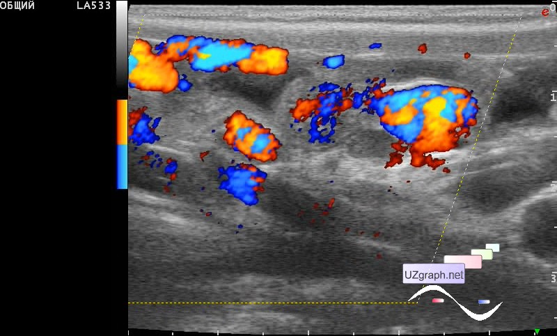
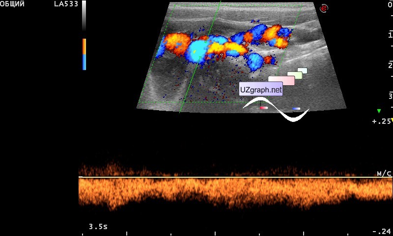
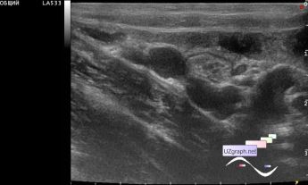
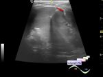
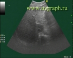
    At ultrasound, the appendix was not visualized, but in the lower right quadrant (RLQ) of the abdomen the tubular convoluted structure was visualized, which I initially interpreted as mesadenitis, on the CFM(DPD) the picture changed, the structure uniformly colored, in the spectral Doppler mode the monophasic blood flow (by venous type) upto 16 cm / sec - presumably phlebectasia/ congenital anomalous blood vessel - external link

    external link

:: attachments(4) ::

:: file 1 ::

:: file 2 ::

:: file 3 ::

:: file 4 ::


 Random 
Ultrasound cases - PDA before surgery
    

On-call room - ARDMS exam

Ultrasound cases - After oncosurgery
    

Technical - Troubleshooting ultrasound scanners

Admin room - New flash-components

 Dictionary 
Anatomical M-mode

LN

IVC

LMP

EFW

LVEDD

EDD

GS

BPD

ESV


 Calculators 
Resistive index

Qp:Qs

Pulmonary artery pressure

TI-RADS

Volume (ellipse formula)


 Similar topics 
Ultrasound cases - Hirschsprung disease on ultrasound
    

Ultrasound cases - Intestinal Infiltrate
    

Ultrasound cases - Abdominal cryptorchidism
    

Ultrasound cases - Diffuse heterogeneous liver
    

Ultrasound cases - Postoperative abdominal wall fistula
    

Ultrasound cases - Stump appendicitis
    

Ultrasound cases - Liver scar after resection
    

Ultrasound cases - Subdiaphragmatic movable fluid collection (UFO)
    

Ultrasound cases - Lesion of the spleen or the case when it is easy to not notice
    

Ultrasound cases - I’m normal but others ...