Log in    RU  


Main page :: Forum :: Ultrasound cases - Sonograms, cine-loops etc. ::

Pneumatosis intestinalis, intussusception etc.


Tags: Gastrointestinal sonography, Esaote MyLab 70, Images, Video, Clinical report, Pediatric

   Posts
 
  Pneumatosis intestinalis, intussusc...
#1

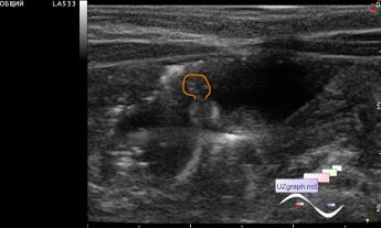
    Newborn with blood in feces and suspected necrotizing enterocolitis was addressed to ultrasound.

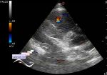
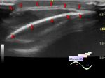
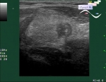
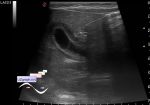
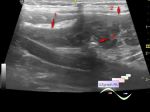
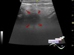
    At ultrasound visualized intestinal loops with hyperechoic dots in the projection of intestinal wall(possible air bubbles in intestinal wall produced by bacteria).

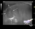
    Also you can see here the target sign - intestinal intussusception, transitory in that case.

    PS. Already during the analysis on the cine-loop in a calm environment(after work), I paid attention to another an/hypoechoic structure(file 5), which was intraluminal and was visible only due to contouring by hyperechoic point inclusions (air bubbles?), and judging by its movement did not allow to exclude the polyp.

    Similar lesion was presented in the another case - https://m.en.uzgraph.ru/forum/...

    A notable moment of such structures(which fixed to the intestinal wall) is that they can be a "lead point" for intestinal intussusceptions.

    external link

:: attachments(5) ::

:: file 1 ::

:: file 2 ::

:: file 3 ::

:: file 4 ::

:: file 5 ::


 Random 
Admin room - Feedback - an error was found

Ultrasound cases - Kidney cystoma
    

Ultrasound cases - Strangulated inguino-scrotal hernia
    

Ultrasound cases - Recurrent shin trauma
    

Ultrasound cases - Femoral lymph node destruction
    

 Dictionary 
SV

MSD

BPD

EFW

LVAs

PW

LMP

B-mode

CSA

LN


 Calculators 
Volume (ellipse formula)

Age / date difference in years and months

Estimated fetal weight by Ultrasound

Modified Simpson’s Rule formula (2 disks) ejection fraction

Type of an infant hip joint


 Similar topics 
Ultrasound cases - Hirschsprung disease on ultrasound
    

Ultrasound cases - Intestinal Infiltrate
    

Ultrasound cases - Subhepatic appendicitis and omentitis
    

Ultrasound cases - Diffuse heterogeneous liver
    

Ultrasound cases - Stump appendicitis
    

Ultrasound cases - Ileostoma stenosis and nephropathy
    

Ultrasound cases - Periappendicular infiltrate
    

Ultrasound cases - Intestinal obstruction, GB wall thickening
    

Ultrasound cases - Ileotiphlitis and mesadenitis
    

Ultrasound cases - Acute Appendicitis, periappendicular infiltrate