Log in    RU  


Main page :: Forum :: Ultrasound cases - Sonograms, cine-loops etc. ::

Intestine contusion


Tags: Gastrointestinal sonography, Esaote MyLab 70, Images, Video, Clinical report, Pediatric

   Posts
 
  Intestine contusion
#1

    A teenager with suspected blunt trauma of the abdomen, is directed to an ultrasound scan of the kidneys with complains on pain in the right half of the abdomen that persists for a day after falling from the bicycle onto the handlebars of the same bicycle.

    Prior to this, during the day, she was already came through the Ambulance to another hospital, where only X-ray was performed, which was without pathology.

    On ultrasound of the abdomen and kidneys - without pathology, but was found abnormal echo-pattern of the part of the intestine, just in the projection of pain - the right upper quadrant of the abdomen.

    In this projection, was visualised a part of the fixed intestine with a thickened wall (6-7 mm) - pseudokidney sign, with a small amount of fluid outwardly surrounded by hyperechoic tissues (omentum? omentitis?), with increased blood flow at DPD - presumably infiltrate within intestinal contusion.

    Emergency hospitalization recommended.

    external link

:: attachments(3) ::

:: file 1 ::

:: file 2 ::

:: file 3 ::

  21:31 10-10-2021
  Ecografía abdominal pediá...
#2

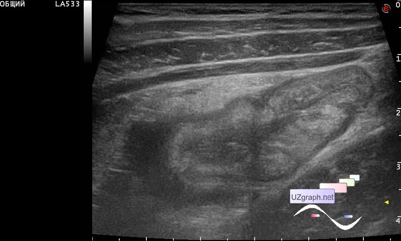
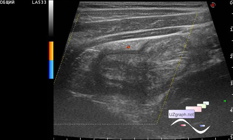
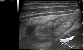
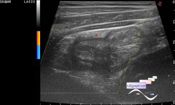
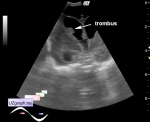
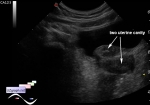
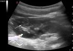
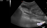
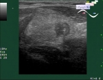
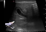
    Una adolescente ingresó en el Hospital Clínico Infantil con un traumatismo abdominal cerrado como consecuencia de una caída de la bicicleta sobre el manillar de la misma bicicleta.

    Previo a esto, durante el día, ya se pasó por la Ambulancia a otro hospital, donde solo se le realizó radiografía, la cual fue sin patología.

    En la ecografía del abdomen y los riñones, sin patología, pero se encontró un patrón de eco anormal de la parte del intestino, solo en la proyección del dolor, el cuadrante superior derecho del abdomen.

    En esta proyección, se visualizó una parte del intestino fijo con una pared engrosada (hasta 6-7 mm / diagnóstico diferido: contusión, hematoma, etc.) - signo de pseudokidney, con una pequeña cantidad de líquido rodeada exteriormente por tejidos hiperecoicos (diagnóstico diferido: infarto omental, omentitis, etc.), con aumento del flujo sanguíneo en DPD - presumiblemente infiltrado dentro de la contusión intestinal.

    external link


 Random 
Ultrasound cases - Submandibular area lesion
    

Ultrasound cases - Abdominal colic
    

Ultrasound cases - Strangulated inguino-scrotal hernia
    

Ultrasound cases - Inguinal-scrotal hernia in infant 2
    

Scientific - Playlist Bones ultrasound

 Dictionary 
FNA

LVOT

IVSd

PG

RVOT

PADP

LVEDV

RAP

VTI

Aliasing doppler artifact


 Calculators 
Volume (ellipse formula)

Teicholz systolic cardiac function

Type of an infant hip joint

Pulmonary artery pressure

Pressure gradient


 Similar topics 
Ultrasound cases - Hirschsprung disease on ultrasound
    

Ultrasound cases - Intestinal Infiltrate
    

Ultrasound cases - Subhepatic appendicitis and omentitis
    

Ultrasound cases - Diffuse heterogeneous liver
    

Ultrasound cases - Stump appendicitis
    

Ultrasound cases - Ileostoma stenosis and nephropathy
    

Ultrasound cases - Periappendicular infiltrate
    

Ultrasound cases - Intestinal obstruction, GB wall thickening
    

Ultrasound cases - Ileotiphlitis and mesadenitis
    

Ultrasound cases - Acute Appendicitis, periappendicular infiltrate