Log in    RU  


Main page :: Forum :: Ultrasound cases - Sonograms, cine-loops etc. ::

Hip dysplasia, subluxation


Tags: Musculoskeletal sonography, Images, Clinical report, GE Logiq P6, Esaote MyLab 70, Pediatric

   Posts
 
  Hip dysplasia, subluxation
#1

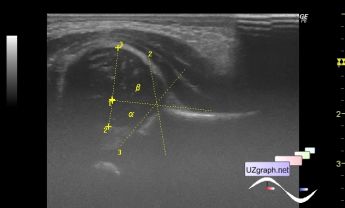
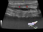
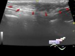
    Newborn with suspicion of hip dysplasia was addressed to ultrasound.

    At ultrasound left hip angles: alpha = 56 deg, beta = 74 deg(Graf type - II), femoral head is lateralized, coverage 33% (Terjesen type - subluxation).

:: attachments(1) ::

:: file 1 ::

  12:37 25-10-2014
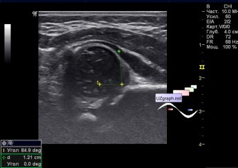
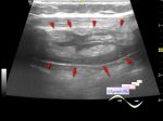
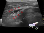
  Another newborn with DDH.
#2

    At ultrasound: left hip angles: alpha = 45-33 deg, beta = 85 deg: Graf type IIc-IV (physiological delay in ossification - Dislocated), femoral head is lateralized, coverage 32% (Terjesen type - subluxation).

:: attachments(2) ::

:: file 1 ::

:: file 2 ::

  04:08 13-05-2015
 
#3

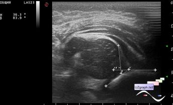
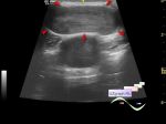
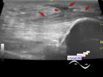
    Another example, a girl 2 months.,

    Graf III-IV(dislocated).

    file 1 - echo

    file 2 - diagram, dotted line shows the normal hip joint(N), regular line - dislocated (D).

:: attachments(2) ::

:: file 1 ::

:: file 2 ::


 Random 
Ultrasound cases - IVSD vs CAF
    

Ultrasound cases - Not a Rubik's Cubes
    

Ultrasound cases - Diffuse-focal thyroid changes, parathyroid glands adenomas
    

Ultrasound cases - Midgut malrotation with volvulus
    

Ultrasound cases - Combined CHD
    

 Ad 


 Dictionary 
M-mode

PI

LVEDV

Aliasing doppler artifact

GS

LVLd

FL

LVAs

SF

Gain


 Calculators 
Pulsatility Index

Area-length (Simpson) formula ejection fraction

Age / date difference in years and months

Thyroid volume

Game: Sonographer certification


 Similar topics 
Ultrasound cases - Penetrating injury of the femur
    

Ultrasound cases - Cephalohematoma
    

Ultrasound cases - Cranial mass
    

Ultrasound cases - Recurrent shin trauma
    

Ultrasound cases - Knee joint synovitis
    

Ultrasound cases - Thickening and heterogeneity of the intercostal space soft tissues
    

Ultrasound cases - Chest wall lesion
    

Ultrasound cases - Hockey Elbow
    

Ultrasound cases - Combination of left shoulder lesions
    

Ultrasound cases - Baker's cyst and intramuscular hematoma
    

 Ad