Log in    RU  


Main page :: Forum :: Ultrasound cases - Sonograms, cine-loops etc. ::

Baker's cyst and intramuscular hematoma


Tags: Musculoskeletal sonography, Soft tissues sonography, GE Logiq E, Clinical report, Pediatric

   Posts
 
  Baker's cyst and intramuscular ...
#1

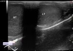
    A 6-year-old girl involved in dancing came to the public clinic for a follow-up ultrasound of Baker's cyst on the right.

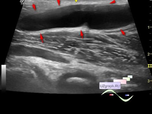
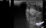
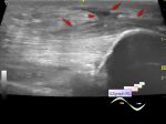
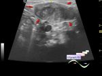
    In the popliteal region on the left, a heterogeneous an-/ hyperechoic inclusion, similar to a complex cyst, without blood flow, without clear boundaries with the muscle, up to 17 x 6 x 21 mm in size is visualized (differential diagnosis: intramuscular hematoma / partial rupture of the muscle, etc. ).

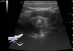
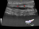
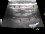
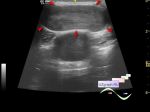
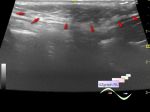
    In the popliteal region on the right, an anechoic inclusion with a clear contour, without blood flow, up to 47 x 9 x 26 mm in size is visualized (differential diagnosis: Baker's cyst, etc.)

    Orthopedic consultation recommended.


    external link


 Random 
Ultrasound cases - Acute phlegmonous appendicitis
    

Ultrasound cases - Choledochal cyst
    

Admin room - Feedback - an error was found

Ultrasound cases - Scapula exostosis
    

Ultrasound cases - Hygromes of wrist
    

 Dictionary 
CFM

PG

EDV

PW

ESV

FNA

PRF

RAP

Gain

PADP


 Calculators 
TI-RADS

Myocardial mass

Qp:Qs

Estimated fetal weight by Ultrasound

Body surface area


 Similar topics 
Ultrasound cases - Penetrating injury of the femur
    

Ultrasound cases - Cephalohematoma
    

Ultrasound cases - Cranial mass
    

Ultrasound cases - Recurrent shin trauma
    

Ultrasound cases - Knee joint synovitis
    

Ultrasound cases - Thickening and heterogeneity of the intercostal space soft tissues
    

Ultrasound cases - Chest wall lesion
    

Ultrasound cases - Hockey Elbow
    

Ultrasound cases - Combination of left shoulder lesions
    

Ultrasound cases - Torticollis / neoplasia